UNIVERSITÀ DEGLI STUDI DI MILANO
|
|
|
- Annabella Parisi
- 8 anni fa
- Visualizzazioni
Transcript
1 UNIVERSITÀ DEGLI STUDI DI MILANO SCUOLA DI DOTTORATO TERRA, AMBIENTE E BIODIVERSITÀ DIPARTIMENTO DI SCIENZE BIOMEDICHE PER LA SALUTE Dottorato di Ricerca in Scienze Naturalistiche e Ambientali Ciclo XXVI-BIO/08 NUOVE TECNOLOGIE NELL AMBITO DELL ANTROPOLOGIA FISICA E FORENSE: IMAGING E MODELLAZIONE 3D Ph.D. Thesis Tutor: Prof.ssa Cristina Cattaneo Daniel Angelo Gaudio Matricola: R09007 Coordinatore: Prof. Nicola Saino Anno Accademico 2012/2013

2 INDICE Indice 1 Capitolo 1 introduzione generale Antropologia fisica e forense, definizione e prospettive d indagine Nuovi tipi di tecnologie al servizio dell antropologo Scopo della ricerca Campi d applicazione Documentazione, profilo biologico, identificazione Lesività scheletrica 11 Capitolo 2 Excavation and study of skeletal remains from a World War I Mass Grave Capitolo 3 Surface curvature of pelvic joints from three laser scanners: separating anatomy from measurement error Capitolo 4 Reliability of cranio-facial superimposition using 3D skull models Capitolo 5 Does cone beam CT actually ameliorate stab wound analysis in bone? Capitolo 6 Application of high resolution pqct analysis to forensic cases for the assessment of bone trauma: a technical note Capitolo 7 The application of cone-beam CT in the aging of bone calluses: a new perspective? Capitolo 8 Indagini preliminari e materiale in elaborazione Age estimation from canine volumes A preliminary study of virtual facial reconstruction by means of 3D models acquired by Laser Scanner and a new facial reconstruction software 105 Conclusioni 114 Appendice 117 1

3 CAPITOLO 1 Introduzione generale 1.1 Antropologia fisica e forense, definizione e prospettive d indagine. Quatrefages de Bréau (Vallerange, Parigi 1892) fu un naturalista francese che si occupò, tra l altro, della dentatura dei roditori, dell anatomia degli anellidi, di molluschi bivalvi e perfino della fecondazione artificiale del pesce. Era un naturalista a tutto campo, uno dei più importanti del diciannovesimo secolo. La ragione per cui egli introduce questa tesi di dottorato è però dovuto al fatto che Quatrefages s interessò anche alla specie umana, al cui studio dedicò buona parte della sua vita. Egli fece chiamare cattedra di antropologia quella di anatomia e storia naturale dell'uomo di cui era titolare; si può dire che la sua fu la prima cattedra ufficiale di antropologia (1855). Definì l antropologia come la storia naturale dell uomo. Tale definizione è sostanzialmente valida anche oggi: secondo l American Association of Physical Anthropology, l Antropologia fisica (o biologica) è definibile come (...) biological science that deals with the adaptations, variability, and evolution of human beings and their living and fossil relatives. Because it studies human biology in the context of human culture and behavior, physical anthropology is also a social science. ( L antropologia fisica è una disciplina con più sotto settori: si parla di paleontologia umana, o paleoantropologia, per indicare lo studio dell evoluzione umana: dalle scimmie antropomorfe, alle prime specie umane fino all unica specie del genere Homo attualmente esistente, l Homo Sapiens. Con Genetica e antropologia delle popolazioni s intende lo studio delle differenze genetiche nelle popolazioni umane. Si parla di antropologia fisica applicata all archeologia (o osteoarcheologia, o anche bioarcheologia) quando ci si riferisce allo studio dei resti umani rinvenuti in siti e contesti archeologici. L antropologia forense, infine, è l applicazione dell antropologia fisica ai casi giudiziari e a quelli riferibili a violazioni dei diritti umani. 2

4 L antropologia forense si occupa, tra l altro, dell identificazione di resti umani scheletrizzati, gravemente compromessi dalla putrefazione o comunque non identificabili (Cattaneo, 2004). Questa tesi di dottorato si occuperà di tematiche e metodiche relative all antropologia fisica e forense. In antropologia fisica e forense il complesso degli strumenti in dotazione all antropologo permettono di ottenere un identificazione generica del soggetto (il suo profilo biologico: sesso, età, statura, etnia) e informazioni circa le patologie di cui il soggetto soffriva. Lo studio di lesività permette di valutare le lesioni perimortem che possono dare indicazioni circa la modalità e la causa di morte. Tali informazioni contribuiscono, in osteoarcheologia, a raccogliere dati circa le caratteristiche, lo stile di vita e lo stato di salute di una popolazione antica. In antropologia forense l analisi dello scheletro fornisce informazioni fondamentali nelle indagini giudiziarie: l antropologo, ove possibile, non si limita all identificazione generica ma fornisce informazioni utili circa l identificazione personale del soggetto. Le operazioni preliminari che l antropologo compie in laboratorio sono il lavaggio delle ossa, il loro restauro (qualora necessario) e quindi lo studio vero e proprio. Pare opportuno indicare quali sono gli strumenti metodologici che l antropologo utilizza nel corso delle indagini. L antropologo studia lo scheletro umano sia macroscopicamente sia microscopicamente. Le metodiche macroscopiche prevedono analisi quantitative (si parla di antropometria, da condurre tramite appositi strumenti di misurazione) e qualitative (metodiche antroposcopiche), che si basano prevalentemente sull osservazione delle ossa. Le analisi microscopiche prevedono l utilizzo di microscopi ottici, stereo microscopi e microscopi elettronici al fine di valutare caratteri o condurre misure non visibili e praticabili a occhio nudo. Lo studio radiologico permette all antropologo di visualizzare dettagli intrinseci agli elementi ossei e dentari. Come vedremo tali dettagli possono essere utili sia per ricavare informazioni circa le affezioni delle ossa, sia per raccogliere informazioni generali relative al soggetto a cui appartiene l osso in esame. Vi sono infine metodiche biomolecolari che si basano sullo studio delle proteine e, naturalmente, del DNA (Cattaneo 2004). All enorme sviluppo in questi decenni delle metodiche biomolecolari, grazie alle recenti innovazioni tecniche (PCR, AMS, Proteomica) e le sempre più efficaci metodologie di estrazione del DNA, sono affiancate nuove forme di tecnologia relative agli altri campi dell antropologia fisica (Kuzminsky, 2012) che, altrettanto velocemente, anche se in maniera meno eclatante, stanno innovando il modo di documentare e d indagare dell antropologo. 3
 e informazioni circa le patologie di cui il soggetto soffriva.")
5 1.2 Nuovi tipi di tecnologie al servizio dell antropologo I metodi di imaging tridimensionale (3D), o diagnostica per immagini tridimensionali, hanno conosciuto un enorme sviluppo in differenti campi negli ultimi decenni; l antropologia fisica (e forense) è una disciplina che si sta giovando di tale sviluppo sia sul piano documentativo, sia sul piano diagnostico. Negli ultimi anni molti laboratori di antropologia hanno iniziato a costituire partnership con scienziati operanti in altri campi, in particolare quello biomedico, al fine di poter utilizzare tecnologie molto costose (ad esempio Tomografia Assiale Computerizzata (TAC), risonanza magnetica (MRI), terahertz imaging (THz), etc.) (Allam et al., 2011; Buikstra, 2010; Conlogue et al., 2008; Faccia and Williams, 2008; Öhrström et al., 2010; Panagiotopoulou, 2009; Saitou et al., 2011) ma di altissimo potenziale ai fini delle indagini antropologiche. Il primo tentativo di imaging su materiale antropologico risale al 1896 (si trattava di uno studio radiografico su mummie umane; Chhem, 2008) ma è con l invenzione delle Tomografia Assiale Computerizzata (nel corso degli anni 70) che si ha il passaggio da una dimensione delle RX alle tre dimensioni dei file DICOM, con la conseguente possibilità di una ricostruzione tridimensionale dei reperti anatomici, una migliore visualizzazione delle superfici e l opportunità di poter condurre analisi accurate anche su corpi non scheletrizzati, difficilmente indagabili se non con metodiche destruenti. Quest ultima possibilità ha dato un decisivo impulso allo studio dei corpi mummificati permettendo di ottenere ricostruzioni virtuali tridimensionali di ossa, tessuti e organi di qualunque epoca, tanto da divenire prassi in questo tipo di indagini antropologiche. Tali strumentazioni, essendo fisse, d altro canto prevedono lo spostamento del materiale d indagine presso le strutture dotate di tali macchine. L antropologo non può condurre le indagini in maniera indipendente, essendo legato a un servizio e al personale tecnico prestato temporaneamente all antropologia, ma la cui funzione originale è prettamente clinico/ospedaliera. L antropologia può tuttavia attingere ad altre tecnologie, fino a qualche anno fa costosissime, ma che stanno divenendo accessibili grazie all abbassamento dei costi e delle dimensioni delle strumentazioni, alla crescente facilità d utilizzo e alla disponibilità di Personal Computer sempre più potenti (in termini di Gigabyte delle schede grafiche, di memoria RAM, di disponibilità di memoria su disco fisso, etc.). Ci si riferisce in particolare alla tecnologia Laser Scanner 3D. Tale tecnologia si è sviluppata in ambito industriale ma ha trovato applicazioni in numerosissimi campi: dall architettura al rilievo dei beni artistici, in ambito archeologico e nelle scienze naturali (ad esempio nei rilievi geologici e paleontologici) fino appunto all antropologia fisica. La scansione laser 3D viene effettuata mediante i Laser Scanners, digitalizzatori ottici che permettono il rilevamento di oggetti e superfici a scale diverse riproducendo dei modelli tridimensionali in 4
, risonanza magnetica (MRI), terahertz imaging (THz), etc.) (Allam et al., 2011; Buikstra, 2010; Conlogue et al.")
6 ambiente virtuale (e del cui principio di funzionamento si accennerà nel secondo capitolo). Il prodotto di una scansione condotta con Laser Scanner è una nuvola di punti georeferenziati (dotati quindi delle tre coordinate spaziali x,y,z) che formano il reperto scansionato. Il modello 3D può anche essere costituito da una griglia (mesh), la quale non è altro che il prodotto dell unione dei punti, che formano quindi una rete di poligoni. La struttura del modello può dipendere dalla tipologia di Laser usata o dal software di elaborazione scelto, è possibile comunque passare da una nuvola di punti a una mesh (e viceversa) mediante un qualunque software di elaborazione 3D. La scansione può essere condotta direttamente in sede di scavo, questo permette di documentare le ossa nel loro contesto originale e acquisire tutte le informazioni morfometriche che caratterizza il luogo di giacenza originale. Si avranno in questo caso nuvole di punti composti da milioni di punti, che dovranno essere opportunamente filtrati ed elaborati. I modelli 3D di reperti ossei sono stati al centro di progetti in ambito paleoantropologici e paleopatologico, dal NESPOS (progetto internazionale di ricostruzione virtuale dei fossili di ominidi), ai progetti Digitized Diseases ( e From Cemetery to Clinic ( e, inoltre, nella digitalizzazione 3D dei reperti ossei della collezione dello Smithsonian Museum. Tra il 2006 e il 2009 la Comunità Europea ha finanziato il progetto EVAN (European Virtual Anthropology Network) con lo scopo di incrementare le metodologie di studio in ambito morfometrico e di varianza anatomica nell antropologia fisica (Sholts et al., 2011). Qualunque sia lo strumento utilizzato per scansionare un reperto scheletrico, una volta ottenuto un modello 3D si può procedere, tramite opportune procedure di conversioni di formati, anche alla stampa tridimensionale del reperto. Si tratta della Stereolitografia, una tecnica che permette di realizzare singoli oggetti (in resina, o materiale termoplastico) a partire direttamente da dati digitali (in termini pratici vi è la concreta possibilità di scansionare un reperto a Tokyo, condividere il file tramite Web, e ottenerne una copia in resina a Milano nel giro di poche ore). Il tema della documentazione e della ripetibilità delle indagini risulta certamente importante in ambito archeologico (e più in generale in ogni ambito scientifico) ma è cruciale nel delicato ambito dell antropologia forense dove, per essere in linea con i dettami relativi agli accertamenti giudiziari, le indagini devono essere non invasive, ripetibili, non modificabili e il più possibile oggettive (Cattaneo, 2004). Appaiono quindi fondamentali tutte quelle tecniche che permettono la documentazione meno invasiva e meno distruttiva possibile dei reperti. Come si accennava, le innovazioni tecnologiche stanno contribuendo al forte sviluppo metodologico nell antropologia, e appare scontato che tali innovazioni possono avere risvolti 5
 mediante un")
7 importanti nei diversi ambiti antropologici; sarebbe tuttavia forviante pensare che esse possano sostituire le metodiche classiche e il giudizio critico dell operatore. Nell ottica dell indagine oggettiva relativa alle potenzialità e ai limiti dell apporto di tale tecnologie, si fonda l approccio con cui è stata condotta questa ricerca. 1.3 Scopo della ricerca Scopo di questa ricerca è quindi quello di indagare le potenzialità e i limiti di nuove tecnologie 3D da utilizzare per la documentazione, l archiviazione e la diagnostica nell ambito dell antropologia fisica e forense. Verificare inoltre l effettivo vantaggio dell informazione che può essere estratta dai dati 3D rispetto alla tipologia di informazioni e documentazione correntemente usata, non tanto per sostituire le metodiche in uso, quanto per integrarle. La ricerca è stata volta in primis a valutare le tecnologie più promettenti in tema di antropologia virtuale; si è sperimentato pertanto l uso di un Laser Scanner 3D di proprietà del Laboratorio di Antropologia e Odontologia Forense (LabAnOF) del Dipartimento di Morfologia Umana dell Università di Milano. In base ai risultati ottenuti e agli ambiti di ricerca indagati si è altresì optato per ulteriori strumentazioni non disponibili presso il LabAnOF; sono state quindi condotte collaborazioni con laboratori e strutture dotate della strumentazione utile allo svolgimento di questa ricerca. Allo scopo di indagare le potenzialità documentative e di archiviazione virtuale 3D è stata acquisita digitalmente la più ampia varietà di materiale antropologico possibile: da reperti ossei sullo scavo, ai singoli elementi scheletrici acquisiti in laboratorio, fino a soggetti viventi. Si è cercato inoltre di studiare le più efficaci modalità tecniche di acquisizione per i diversi tipi di materiali e soggetti, verificando le problematiche legate a limiti di scansione in base alle caratteristiche intrinseche del materiale acquisito e al tipo di strumento utilizzato. Sono state valutate quindi le procedure di elaborazione digitale dei modelli ottenuti sia ai fini forensi, sia a quelli osteoarcheologici. Sono stati infine studiati e applicati numerosi software per l elaborazione digitale, ricercando inoltre i formati digitali più idonei all archiviazione dei dati al loro trasferimento su più programmi. Sono state poi selezionate alcune problematiche di antropologia fisica e forense che da un lato necessitano di miglioramento metodologico e diagnostico, da ricercare anche mediante l utilizzo delle nuove tecniche d acquisizione e modellazione tridimensionale, dall altro che rispondono alle concrete esigenze investigative di un laboratorio di Antropologia Forense impegnato nella soluzione di casi di natura giudiziaria. 6

8 La documentazione, acquisizione e archiviazione dei dati tridimensionali, ha reso possibile, tramite l utilizzo delle tecnologie 3D, un indagine relativa alla diagnosi d età e alla ricostruzione facciale, argomenti che rientrano, in antropologia, nella stesura del profilo biologico. L uso del modello 3D è stato applicato alla tecnica di sovrapposizione cranio-facciale (che rientra nei metodi d identificazione personale). Si è inoltre affrontato il tema della lesività scheletrica, verificando la capacità documentativa e diagnostica delle lesioni antemortem (calli ossei) e della lesività perimortem mediante metodologie di esplorazione e ricostruzione delle lesioni in ambiente virtuale. 1.4 Campi d applicazione Documentazione, profilo biologico, identificazione. Una delle operazioni fondamentali nell indagine antropologica è la documentazione dei reperti che si stanno indagando. Fondamentale nell ambito archeologico, sia in sede di scavo, per l appropriata documentazione dello stesso, sia dei singoli reperti poiché una volta studiati solitamente sono archiviati e/o restituiti al committente dello studio. Il materiale oggetto di indagine deve quindi essere ben documentato sia per offrire un efficace supporto alla relazione antropologica, sia perché potrebbe non essere più a disposizione dell operatore una volta concluso lo studio. In ambito forense, oltre alle motivazioni appena esposte, l adeguata documentazione è cruciale poiché, se non condotta sotto il corretto profilo giudiziario, può non solo inficiare l indagine ma anche un intero processo (Cattaneo, 2004). L avvento della fotografia digitale ha rivoluzionato il mondo del rilievo fotografico, rendendo possibile la produzione di enormi quantità di fotografie ad alta risoluzione scaricabili su un qualunque disco, fisso o removibile, a sua volta sempre più capiente. Erano solo i primi anni novanta quando i modelli di fotocamera digitale più all avanguardia raggiungevano la risoluzione di 1,5 MP (MegaPixel). Un modello della Kodak (DCS200) con tali caratteristiche costava circa dollari (Reis, 2008). Oggi per meno di 30 euro ognuno di noi può acquistare una fotocamera digitale a 3 MP. Un laboratorio può investire circa 100 euro per comprare una fotocamera da 12 MP, dotata di funzione Macro, che permette di fotografare oggetti a pochi centimetri di distanza dall'obiettivo (ideale per ritrarre i dettagli), ottenendo immagini di qualità impensabile fino a pochi anni fa. Le fotografie e il rilievo fotografico in genere hanno tuttavia delle difettosità intrinseche : 7
 e della lesività perimortem mediante metodologie")
9 Bidimensionalità: una fotografia rappresenta quanto catturato su un asse X e un asse Y. Questa caratteristica rappresenta il principale limite; una foto non consente di discernere perfettamente i livelli di profondità di un dettaglio (ad esempio di una lesione). Difficoltà nel legare il generale al particolare: in altri termini se una foto a forte ingrandimento rende bene i particolari, il particolare perde parzialmente la sua collocazione nel contesto generale (principio di indeterminazione). Impossibilità di misurazione: una foto bidimensionale non consente la misura tra due punti, ma solo la stima in base all inserimento nel campo fotografico di un repere metrico di riferimento (il classico righello o meglio la scala ABFO). Tali difettosità intrinseche delle immagini 2D rendono ragione della differenza tra visione diretta e resa fotografica, infatti il nostro occhio è strutturato per la visione tridimensionale e i livelli di profondità contribuiscono in modo fondamentale alla definizione della visione. Il contributo della tecnologia 3D alla documentazione volge appunto al superamento di questi limiti. Il processo di digitalizzazione di un oggetto fisico per l'analisi o la rimodellazione computerizzata di superfici geometriche, mediante apposite attrezzature e rielaborazioni, si sviluppa in due fasi: la prima è denominata fase di acquisizione dei dati; la seconda è la fase di elaborazione degli stessi. I sensori tridimensionali (Laser Scanner) sono strumenti che permettono di generare un immagine 3D dell oggetto che inquadrano. A differenza della tradizionale fotografia, sono in grado di descrivere l'oggetto rilevato nelle sue tre dimensioni spaziali riuscendone a restituire una misura oggettivizzata. In generale, si distinguono due principali famiglie di scanner: i ranging scanners (scanner a misura diretta dalle distanza, tra cui quelli a tempo di volo) ed i triangulation scanners (scanner a triangolazione). Nei primi, la posizione dell emettitore laser e del ricevitore coincidono. Nei secondi, emettitore e ricevitore sono separati da una distanza nota a priori (base line) sulla quale si basa il principio della triangolazione. Possono acquisire la superficie esterna della scena e dell oggetto, ma non possono penetrare sotto le superfici. (Sgrenzaroli 2013). La tomografia assiale computerizzata, nata dall esigenza di indagare le strutture interne di un elemento anatomico, restituisce anch essa un modello tridimensionale dell oggetto acquisito. Un modello 3D è una rappresentazione digitale, fedele e misurabile, dell oggetto in esame, ottenuta mediante la rappresentazione esplicita delle sue caratteristiche morfometriche. Alcuni autori hanno confrontato metodi diversi per la produzione di modelli tridimensionali e valutato l accuratezza nelle misure condotte sui modelli ottenuti. Fourie (2011) ha testato l accuratezza del Laser Scanner Minolta Vivid 900 rispetto a una Tac Cone Beam, scansionando dei volti e rilevando l estrema accuratezza (sub millimetrica) di entrambi gli strumenti. 8
.")
10 Il presente studio ha altresì indagato i vantaggi e gli svantaggi dell utilizzo del Laser Scanner rispetto alle TAC di nuova generazione (in particolare CBCT e pqct) nel tentativo di individuare lo strumento più adatto da utilizzare in relazione alle caratteristiche del reperto, al tipo di documentazione da condurre, e all obiettivo della documentazione stessa. All acquisizione del modello, come si diceva, segue una fase di elaborazione del dato 3D tramite appositi software; l elaborazione prevede l importazione del dato 3D all interno del software, eventualmente il filtraggio dei dati ridondanti (rumore), l editing dei dati, il mashing (l eventuale creazione di una rete poligonale dei dati), e infine l esportazione del dato 3D attraverso la conversione nel formato che si ritiene più idoneo. Nel presente studio si è verificato la capacità documentativa di un Laser Scanner a tempo di volo nel documentare uno scavo archeologico, in particolare una fossa comune risalente alla Grande Guerra (Capitolo 2). Spostandosi dallo scavo al laboratorio si è testata la riproducibilità di superfici articolari di sinfisi pubiche e superfici auricolari dell ileo, acquisite con tre differenti laser scanners a triangolazione ottica (adatti quindi a reperti di piccolo e medie dimensioni) (Capitolo 3). In conseguenza dell ottenimento di un modello tridimensionale è possibile procedere in diversi ambiti dell antropologia fisica e forense, nel corso di questo studio si è indagato in particolare: - La stesura del profilo biologico: consiste nella raccolta, da parte dell antropologo, di tutti i dati informativi che possono essere desunti dallo studio dei resti scheletrici relativamente al sesso, l età, la razza, la statura e, eventualmente, le possibili caratteristiche fisiognomiche del soggetto. In ambito osteoarcheologico la stesura del profilo biologico permette non solo la raccolta dei caratteri di uno scheletro ma anche di raccogliere informazioni utili a definire la struttura biodemografica della popolazione antica cui appartiene (Canci 2005). In ambito forense la stesura del profilo biologico di un soggetto può circoscrivere la cerchia dei sospetti d identità nel caso di un individuo non identificato, oppure avvalorare o smentire l ipotesi identificativa su un soggetto rinvenuto in un contesto critico (si pensi ad esempio a un soggetto carbonizzato in auto). Il profilo biologico risulta inoltre fondamentale per distinguere persone diverse in contesti di ossa commiste (Adams and Byrd 2006) e nei disastri di massa. In particolare è stata condotto uno studio circa l utilizzo dell imaging 3D per la diagnosi d età a partire dall estrazione di volumi dentari (capitolo 8.1): la valutazione dell'età biologica è una disciplina in continua evoluzione che trova le sue principali applicazioni in ambito clinico (diagnosi, prognosi e terapia auxologiche e odontoiatriche) e in ambito forense relativamente alla stima dell età cronologica a essa correlata sia su cadaveri sia su viventi. 9
, l editing dei dati, il mashing (l eventuale creazione di una rete poligonale dei dati), e infine l esportazione del dato 3D attraverso la")
11 Ci si è inoltre rivolti al tema della ricostruzione facciale (capitolo 8.2), anch esso rientrante nella ricostruzione del profilo biologico. La ricostruzione facciale è l operazione di riproduzione di un volto a partire da un cranio. Nel corso di questa ricerca si è valutato l utilizzo del Laser Scanner 3D rispetto alla TAC tradizionale nell acquisizione di crani sui cui condurre la ricostruzione facciale; si è inoltre operata la ricostruzione facciale utilizzando un nuovo software per la ricostruzione facciale (AFA 3D TIVMI) che opera applicando automaticamente un numero molto elevato di spessori tissutali (78), con origine in altrettanti punti craniometrici, ottenuti mediante una ricerca di valori tissutali condotti su una vastissima popolazione europea (500 individui). Ci si è poi rivolti a un particolare e delicato ambito dell antropologia, quello dell identificazione personale. Accertare l identità da cadaveri o resti umani o resti scheletrici consiste nel riconoscere i caratteri individuali che differenziano inconfondibilmente un determinato individuo da qualsiasi altro individuo (Cattaneo 2004). È un argomento che inserito nelle ricerche antropologiche riguarda prevalentemente (ma non esclusivamente) l antropologia forense. L identificazione personale, per definirsi tale, viene basata sulla cosiddetta physical evidence, che si può ottenere attraverso indagini genetiche, dattiloscopiche, odontologiche e osteologiche. Vi possono essere casi in cui le tecniche più diffuse d identificazione non possono essere condotte: si pensi ad esempio al rinvenimento di resti umani di immigrati, di cui non si ha notizia di parenti (cosa che purtroppo accade spesso) e in cui non è possibile avere confronti digitali per compromissione dei polpastrelli a causa dei fenomeni decompositivi. Si devono allora utilizzare altri metodi d identificazione. Le tecniche osteologiche prevedono l utilizzo di confronti radiografici dei dati ante/post-mortem, è pertanto necessario poter disporre di documentazione radiografica del soggetto in vita. Oltre alla difficoltà di reperimento del materiale radiografico, l identificazione osteologica è difficilmente quantificabile (al contrario di quella dattiloscopica e genetica) e in letteratura vi è ancora un certo dibattito su come considerare un carattere o forma unica e specifica. In assenza di altre opzioni rimane un ultima tecnica, la sovrapposizione cranio facciale. A patto di possedere una qualunque immagine del soggetto in vita. Tale metodica confronta la morfologia del cranio con la foto del volto di un soggetto scomparso, valutandone la corrispondenza secondo specifici caratteri. Si è condotto uno studio (capitolo 4) per valutare i vantaggi e l affidabilità della tecnica a partire da un modello 3D ottenuto da un cranio scansionato con Laser Scanner. 10
 che opera applicando automaticamente un numero molto elevato di spessori tissutali (78), con origine")
12 1.4.2 Lesività scheletrica Particolare importanza assume la possibilità di documentare lesioni, anche sub millimetriche, presenti nel tessuto osseo. Attraverso le tecniche d acquisizione tridimensionale delle lesioni, la documentazione può divenire metodo che consente l esplorazione delle stesse, e avere, in altre parole, valenze diagnostiche. Sono stati condotti studi relativamente alle lesioni perimortem e antemortem. Lesività perimortem Di particolare importanza, in antropologia forense, sono le lesioni perimortem, cioè quelle lesioni che hanno causato la morte del soggetto o sono dovute a un trauma verificatosi immediatamente prima o dopo la morte del soggetto. A seconda della modalità traumatica si distingue principalmente (si riporta qui solo la lesività che interessa la presente ricerca) una lesività da arma bianca, da arma da fuoco, contusiva e lacerocontusiva. Lesività da arma bianca (Cattaneo, 2004; Arbarelo,1999): a seconda del tipo di arma bianca avremo differenti tipi di lesione: - lesioni da punta: nell osso e nella cartilagine, data la poca elasticità di questi tessuti, sono caratterizzate da fori, a fondo cieco o trapassanti, che conservano la forma e le dimensioni della punta feritrice. - lesioni da taglio: raramente sono penetranti (interessano prevalentemente parti molli); si possono però avere intaccature delle ossa, di regola superficiali ma talora anche resecanti. Quando il filo della lama scorre sull osso produce fessurazioni che si presentano con conformazione a V; agli estremi della lesione possiamo rinvenire le cosiddette codette, segni prodotti dal filo della lama che via via si riporta verso la superficie. - lesioni da punta e taglio: ferite di questo tipo vengono generate quando la lama presenta un estremo acuminato e uno o più spigoli affilati taglienti; variano di aspetto a seconda che la lama sia monotagliente, bitagliente, tritagliente o tetratagliente. Le ferite d arma da taglio, e in particolare da coltello, sono fra le più frequenti cause di morte nei casi di omicidio (Martin 1999).Gli studi qui presentati (capitoli 5 e 6) si sono in particolare focalizzati alla documentazione e analisi di questo tipo di lesività. Lesività antemortem 11

13 Quando un trauma genera lesioni dell osso sulla persona in vita, provocando una frattura parziale o totale dell osso stesso ma non la morte dell individuo, il processo di guarigione comporta la formazione di un callo osseo che è conseguenza di una risposta di tipo proliferativo da parte dell osso leso; in seguito avviene il rimodellamento e l arrotondamento con formazione di osso lamellare nel tratto danneggiato. Il callo osseo costituisce la fase ultima del processo di guarigione di una frattura, con la fusione dei due monconi ossei dovuta alla deposizione di osso neoformato. La guarigione può essere completa o, a seconda del trauma subito, portare a una menomazione residua del segmento scheletrico. In antropologia fisica riconoscere lesioni ante mortem e individuarne le caratteristiche può fornire informazioni utili circa l occupazione e gli stress meccanici a cui l individuo era sottoposto. In ambito forense la possibilità di avere più informazioni circa l epoca di cagione e lo stadio di guarigione di una frattura può supportare le indagini identificative. Se un incremento delle informazioni utili alla datazione dei calli ossei può essere ricavato dalle nuove tecnologie digitali viene indagato e discusso nel capitolo 7. 12

14 Bibliografia Adams BJ, Byrd JE, (2006). Resolution of small-scale commingling: A case report from the Vietnam War', Forensic Science International, 156, Allam AH, Thompson RC, Wann LS, Miyamoto MI, el-halim Nur el-din A, el-maksoud GA, Al-Tohamy Soliman M, Badr I, el-rahman Amer HA, Sutherland ML, Sutherland JD, Thomas GS, (2011). Atherosclerosis in ancient Egyptian mummies: the Horus study. Journal of the American College of Cardiology 4: Arbarelo P, (1999). Compendio di medicina legale, Minerva Medica, Torino. Buikstra, JE, (2010). Paleopathology: a contemporary perspective. In: Larsen, C.S.n A Companion to Biological Anthropology. Wiley-Blackwell, Chinchester. Canci A, Minozzi S, (2006). Archeologia dei resti umani. Carocci, Roma. Cattaneo C, Grandi M,(2004). Antropologia e Odontologia Forense. Guida allo studio dei resti umani. Testo atlante. Monduzzi Editore, Bologna. Chhem RK, (2008). Paleoradiology: History and New Developments. In: Brothwell R, Chhem RK, Paleoradiology, Springer-Verlag Berlin, Heidelberg. Conlogue G, Beckett R, Bailey Y, Posh J, Henderson D, Double G, King, T, (2008). Paleoimaging: the use of radiography, magnetic resonance, and endoscopy to examine mummified remains. Journal of Radiology Nursing 27: Faccia KJ, Williams RC, (2008). Schmorl s nodes: clinical significance and implications for the bioarchaeological record. International Journal of Osteoarchaeology 18: Fourie Z, Damstra J, Gerrits PO, Ren Y, (2011). Evaluation of anthropometric accuracy and reliability using different three-dimensional scanning systems. Forensic Science International 207: Martin JR, (1999). Identifying Osseous Cut Mark Morphology for Common Serrated Knives. Thesis presented for the Master of Art degree, Department of Anthropology, University of Tennessee Knoxville 13
. Compendio di medicina legale, Minerva Medica, Torino.")
15 Kuzminsky SC, Gardiner MS, (2012). Three-dimensional laser scanning: potential uses for museum conservation and scientific research. Journal of Archaeological Science 39: Öhrström L, Bitzer A, Walther M, Rühli FJ, (2010). Technical note: terahertz imaging of ancient mummies and bone. American Journal of Physical Anthropology 142: Panagiotopoulou O, (2009). Finite element analysis (FEA): applying an engineering method to functional morphology in anthropology and human biology. Annals of Human Biology 36: Quatrehomme G., Iscan MY (1999). Characteristics of gunshot wounds in the skull. Journal of Forensic Science 44: Reis G., (2007). Analisi Forense con Photoshop. Apogeo, Lavis (Trento). Saitou N, Kimura R, Fukase H, Yogi A, Murayama S, Ishida H, (2011). Advanced CT images reveal nonmetric cranial variations in living humans. Anthropological Science 119: Sgrenzaroli M, Vassena G, Galassi A, Gaudio D, Martini P, (2013). La rappresentazione della scena del crimine: dalla descrizione narrativa ai rilievi tridimensionali. In: Curtotti D, Saravo L, Manuale delle investigazioni sulla scena del crimine. Giappichelli Editore, Torino. Sholts SB, Flores L, Walker PL, Warmlander SKTS, (2011). Comparison of Coordinate Measurement Precision of Different Landmark Types on Human Crania Using a 3D Laser Scanner and a 3D Digitiser: Implications for Applications of Digital Morphometrics. International Journal of Osteoarchaeology. 21(5):
. Analisi Forense con Photoshop. Apogeo, Lavis (Trento).")
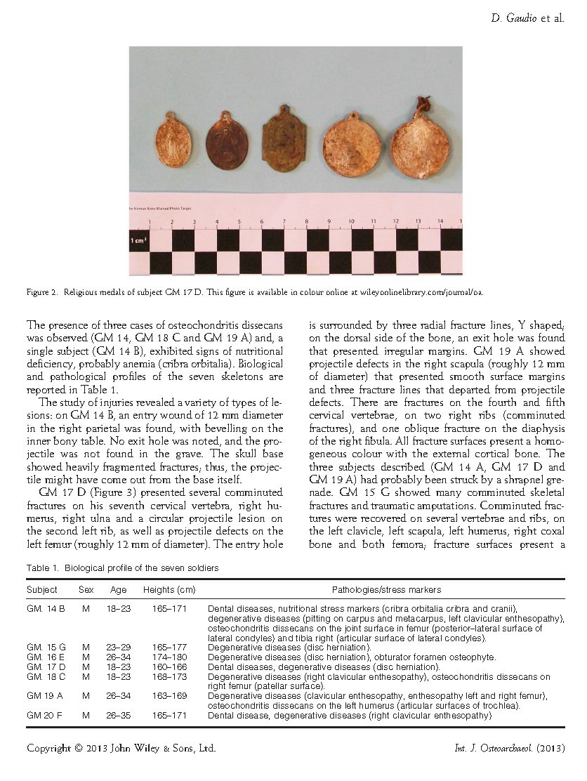
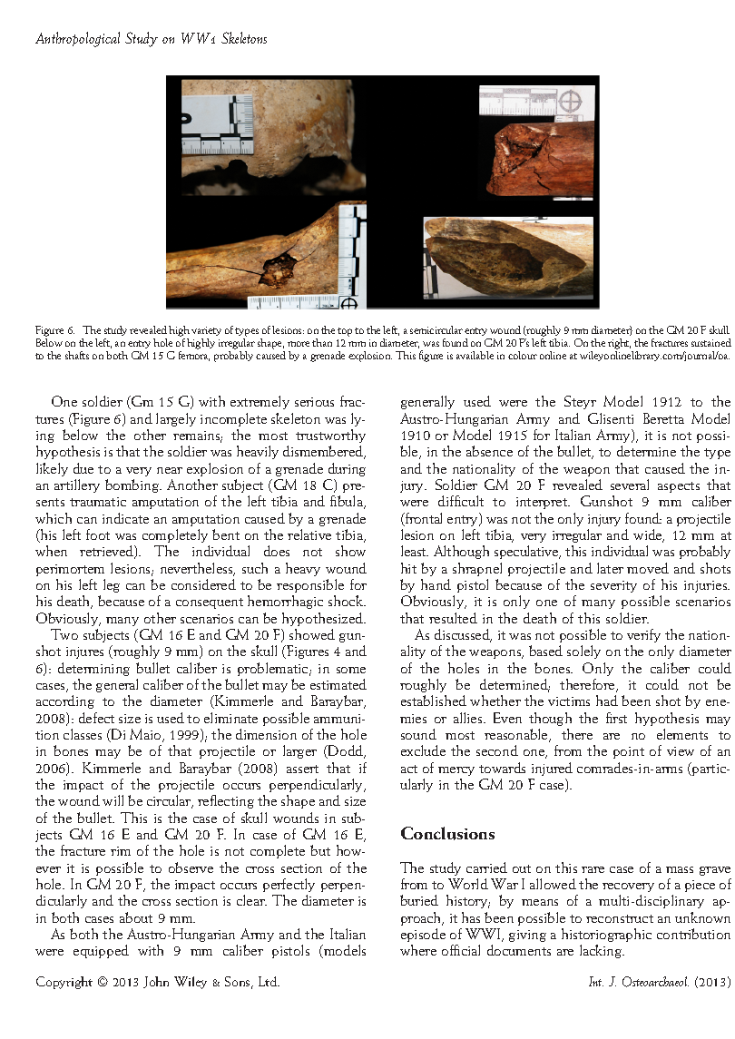
16 CAPITOLO 2 Excavation and study of skeletal remains from a World War I Mass Grave. Gaudio D, Betto A, Vanin S, De Guio A, Galassi A, Cattaneo C. Article first published online: 8 AUG 2013 DOI: /oa

17 16

18 17

19 18
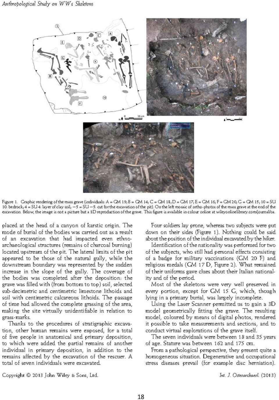
20 19


21 20

22 21
23 22
24 23
25 CAPITOLO 3 Surface curvature of pelvic joints from three laser scanners: separating anatomy from measurement error. Villa C, Gaudio D, Cattaneo C, Buckberry J, Wilson AW, Lynnerup N. Submitted to Journal of Forensic Science 24
26 Surface curvature of pelvic joints from three laser scanners: separating anatomy from measurement error. Chiara Villa 1*, Daniel Gaudio 2, Cristina Cattaneo 2, Jo Buckberry 3, Andrew S.Wilson 3, Niels Lynnerup 1 1 Laboratory of Biological Anthropology, Department of Forensic Medicine, University of Copenhagen, Denmark; 2 LABANOF, Forensic Anthropology and Odontology Laboratory, Department of Human Morphology, University of Milan, Italy. 3 Biological Anthropology Research Centre, Archaeological Sciences, University of Bradford, UK; Keywords: forensic science, forensic anthropology, pubic symphysis, auricular surface, curvature, laser scanner, surface area, distance deviation, 3D models Abstract Recent studies have reported that quantifying symphyseal and auricular surfaces curvature changes on 3D models acquired by laser scanners have a potential for age-at-death estimation. However, no tests have been carried out to evaluate the repeatability of the results between different laser scanners. We calculated and compared the surface curvature of 3D models of symphyseal and auricular surfaces generated from three different laser scanners. The surface area and the distance between co-registered meshes were also investigated. Close results were found for surface areas (differences between 0.3% and 2.4%) and for distance deviations (average < 20 μm, SD < 200 μm). The curvature values were found to be systematically biased between different laser scanners, but still showing similar trends with increasing phases / scores. We filtered the 3D models to separate anatomy from the measurement error of each instrument, so that similar curvature values could be obtained (p < 0.05) irrespective of the specific laser scanner. 25
27 Introduction Recent studies have shown the benefits of quantitative methods using 3D laser scanner models in addressing fundamental issues in physical and forensic anthropology. Sexual dimorphism and population and ancestry variation have been investigated quantifying surface areas or extracting curves [1-5]. The morphological features of the symphyseal and the auricular surfaces used for age estimation have been examined looking at the surface curvature changes [6-8]. Laser scanners have also been used to investigate cranial facial variation and for facial identification [9-13]. In all these applications, the precision and the repeatability of the measurements among different instruments are essential for the reliability of each method; in fact, there are different models of laser scanners and differences between the software used for post-processing the scans (i.e. aligning, merging and fusion of the single scans to create a 3D model). It has been demonstrated that the surface areas were reproduced with high precision and the measurement errors of the extracted information varied from 0.2% to about 1% [5, 14]; it has been also shown that the location of points on the surface (landmarks) and the measurements of length could be accurately repeated on 3D models [11, 12, 15]. When some of the parameters used for the scans made by the same laser scanner have been changed, measurement error was reported to increase slightly to 2% [14]. However, all these studies have tested the repeatability of measurements made by the same instrument. Laser surface scanning (and CTscanning) of bones is proposed as a method to make osteological data more widely and easily available, e.g. the Smithsonian 3D collection [16], "Digitised Diseases [17] and From Cemetery to Clinic [18]. It is thus important to investigate whether 3D models acquired by different laser scanners may exhibit larger differences than those acquired by the same scanner. Irrespective of the 26
28 performance of each laser scanner, the nodes of the resulting 3D model surfaces represent the true anatomical shape, plus some error due to measurement uncertainty. The formal may not always be available, such as calibration steps followed and the exact distance between object and scanner [19, 20]. A simple but general approach to assess measurement error that does not rely on any external knowledge beyond the scanned 3D mesh would therefore be particularly convenient. We assumed the measurement uncertainty manifests itself exclusively as independent random error in the position of expression of this uncertainty is difficult, as it requires full knowledge not only of the instrument and object properties, but also of operational details which generated with three different laser scanners, three different kinds of post-processing software, and the same algorithm used in Villa et al. [6]. To approximate the conditions of an independent replication of this experiment, only the final 3D models were compared, excluding other influences such as the resolution of the instrument, the operational conditions or type of algorithm used to merge the scans. mesh nodes, which can be effectively reduced by a smoothing operation over neighboring Materials and methods nodes. If this assumption is appropriate, the Sample use of an adequate amount of smoothing would filter out the measurement error while preserving the overall anatomical shape of the bone surface, and homogenize 3D models from different instruments. The aim of this paper was to experimentally verify the validity of our assumption by using 3D models of symphyseal and auricular surfaces The sample consisted of the 24 Suchey-Brooks pubic bone casts (12 females, 12 males) [21] and 19 archeological auricular surfaces of the recording kit collected by Buckberry and Chamberlain as illustrative of the different scores of the features described in their method [22]. We selected only the central area of the pubic symphyseal face 27
29 inside the margins and the internal area of the the contour of the joint, as described in [6]. auricular surface, i.e. the area delimitated by Laser scanning Three different laser scanners were used in this study (Table 1): FaroArm Quantum with V3 Laser Line Probe - FARO Singapore Pte. Ltd (abbreviated Faro), property of the University of Bradford (UK); Minolta VI Konica Minolta Sensing, Inc. Osaka-Japan (abbreviated Minolta), property of the University of Milan (Italy); and a custom laser scanner (abbreviated Custom) property of the University of Copenhagen (Denmark) [6]. The Faro is equipped with an articulated arm, thus the object to scan was kept fixed during the scanning process; for the other two laser scanners, the scanning system was fixed while the object was moved. In order to capture as much detail as possible, the object was located as close as the laser scanner allowed. Three different software applications were used for the post-processing: PolyWorks [23] was the software accompanying the Faro instrument, Geomagic Studio [24] was used for the scans from Minolta and David-laser scanner software [25] for those from Custom. In each software, the followed steps were carried out: first, each mesh was visually inspected, and occasional spikes were removed and small holes were filled; second, the individual meshes were aligned (the Faro roughly aligned the individual scans during the scan processing, so a second alignment was performed in this case); third, the aligned meshes were merged for generating the final mesh and removing redundant data; finally, a slight smoothing (the lowest possible smooth allowed by each software) was applied at the merged mesh. The final model was saved in STL format (Standard Triangulation Language) and used in all analysis. Many factors can have an effect on the scans and are difficult to control and reproduce. First, the performance of the different laser scanners rarely can be 28
30 compared since there are no protocols and the precision and accuracy specified by different manufacturers are measured and expressed in different ways [26]. Furthermore, the quality of the resulting 3D models depends not only on the nominal resolution of the laser scanner, but also, among other factors, on the ambient light, the manual skill of the operator ( more evident in Faro), surface color and the geometry of the object, the distance of the scanner to the object and the instantaneous incident angle of the laser and the line of sight of the camera [19, 20]. We were only 3D, so the scans were acquired under optimal conditions by qualified operators. While this may not be the most formally rigorous experimental protocol, it can be argued that it provides the most relevant common ground for comparison among instruments under operational circumstances as close as possible to the real world application of these instruments. As we are only concerned with the practical usability of the typical scans a user will obtain, we have focused on comparing the final 3D models, without emphasizing these other factors further. interested on the differences among the final Portability Weight Texture Velocity of scan (*) FARO Yes ~10 kg No Fast (less than 5) MINOLTA Yes ~11 kg Yes, color Fast (less than 5) Type of laser scanner system Mobile (object fixes) Fixed (object moves) Accuracy mm (a) b X: ±0.22mm Y: ±0.16mm Z: ±0.10mm (b) Type of mesh Triangle, unstructur ed Triangle, structured Range of price ( ) > > CUSTOM Yes ~5 kg Yes, grey Very slow Fixed Triangle, <5 000 scale ( ~15 min) (object mm structured moves) (c) Table 1: Performance parameters of the laser scanners. The accuracy values are taken from the available manufacturers literature. They are not always comparable across instruments and with the actual measurement conditions used in this work (* to scan a pubic bone, (a) volumetric maximum deviation at 1.8m, (b) to the Z reference plane -Conditions: TELE/FINE mode, Konica Minolta s standard, (c) calculated as 0.5% of the object size) 29
31 Surface curvatures, surface areas and distances. For each specimen scanned by each laser scanner, we calculated the mean of the curvature values (as in [6]): therefore, we calculated the curvature of 57 threedimensional models of auricular surfaces (19 specimens scanned three times) and 72 threedimensional models of pubic bones, (24 casts scanned three times). The differences between the 3D models of the same specimen were calculated and expressed in percent. The curvature values of 3D models from Custom laser scanner were used as reference models. To reduce the total curvature differences to a maximum of 2%, we applied a smoothing factor, i.e. we modified the algorithm for calculating curvature to eliminate the To enable a comparison with other studies reported in the literature, we quantified the surface areas (reported in cm 2 ); we also calculated the differences of surface area (expressed in cm 2 and percent) between the 3D models of the different instruments. In additional, we calculated the distance deviation (reported in μm), i.e. the distance between the points of a 3D model of one laser and corresponding ones of second laser scanner. We used a function of Geomagic Studio Software, called deviation, that returned for each two-by-two comparison the Average distance and the Standard Deviation distance ; furthermore this function generated color-coded mapping of the differences, highlighting the areas and the specimens with problems. measurement error of the lasers. 30
32 laser scanner: Faro, Minolta and Custom. Statistical analyses All descriptive statistics (means, standard deviations (SD), and differences) were calculated using SPSS software, version 20. We used scatter plots to undertake a visual evaluation of the distances between 3D models from different laser scanners. Each scatter plot shows the comparison between two laser scanners: 1) Faro versus Minolta; 2) Custom versus Faro; 3) Custom versus Minolta. To highlight any variation with increasing age, we ordered the auricular surface features based on increasing score; the features were used in the following order: TO= transverse organization, ST= surface texture, MA= macroporosity, MI= microporosity, AC= apical change. In the same way, we ordered the pubic bones based on increasing phase; two values were present for each phase, representing the early (E) and the advanced (A) patters. The scatter plots were also used to investigate the curvature values, with the same organization described above; in this case, each series represented a Finally, we performed Kruskal-Wallis ANOVA to compare the revised curvature values, after the use of the smoothing. Results Surface curvature The curvature values of the auricular surface and the pubic bone (male and females separately) are shown in Figure 1. The scatter plots in the column "a" (on the left) show the results obtained using the unmodified algorithm used in Villa et al. [6], with the same smoothing factor for all laser scanners. The curvature values show similar trends with increasing phases and scores: the curvature increases and decreases in the same manner in all three laser scanners, but with a systematic difference among the models of laser scanners. Faro shows the highest curvature values in all samples, both for the pubic bones and the auricular surfaces. Minolta and Custom have closer values, but more than 50% of the curvature values of Minolta are higher than those from Custom, especially in 31
33 the auricular surfaces. In the pubic bones, differences of 28.8% (females) and 20.5% ( males) were found between Faro and Custom, 0.29% (females) and 1.6% (males) between Custom and Minolta. Differences of 44.2% and 9.7% were found in the auricular surfaces, between Faro and Custom, and between Minolta and Custom respectively. To reduce these curvature differences, we modified the amount of smoothing in the curvature algorithm. The curvatures of Faro could be homogenized with Custom using a smoothing factor of 10 for the male pubic bones, of 15 for the female pubic bones, and b ); in this manner, the distance between the two lasers were reduced to about 1.5%. The curvature values of Minolta and Custom in the pubic bones for both sexes were already very close, with a difference lower than 2%, so no further smoothing factor was applied. A smoothing factor of 5 was required to reduce the difference between the auricular surfaces of Minolta and Custom to 1.8%. Figure 1b (scatter plots on the right) show the corrected curvature values. We found no statistically significant difference (p> 0.05) between the corrected curvatures values produced by the different laser scanners. 20 for the auricular surfaces (Fig. 1, column 32
34 Figure 1: Curvature values of the auricular surface (top row) and of the pubic bones (middle row females, bottom row males). Column a shows the curvatures value with the same "smoothing factor" for 3D models by all laser scanners; Column b shows the curvature values with different "smoothing factor" (reported in the legends) 33
35 Surface areas The measurements of the surface area and differences between the laser scanners for the auricular surfaces and the symphyseal surface (males and females separately) are reported in Table 2. The largest difference for the auricular surfaces was 0.15 cm 2 and was found between Faro and Minolta corresponding to 1.5%. In the symphyseal surface, Custom and Faro have the highest differences, cm 2 (2.4 %) in females and cm 2 (1.9%) in males. Surface area (cm 2 ) Area difference (cm 2 ) N Custom Faro Minolta Custom- Faro % Custom- Minolta % Faro- Minolta % Auricular surfaces Pubic bones Females Pubic bones Males ± ± ± % % % ± ± ± % % % ± ± ± % % % Table 2: Mean value and standard deviation of the surface area of the auricular surfaces and the pubic bones for each laser scanner and area differences in cm and percent Distance deviations Figure 2 show the distributions of the "average distances" (black solid points) and average plus/ minus the SD for the auricular two laser scanners. Faro and Minolta show the lowest SD distance, less than 100 μm with a mean average distance of 1.1 μm (Table 3). surfaces. It is clear that there is more variation in distances (most evident in the SD) in the last scores where the surface topography is more irregular: score 3 for AC, scores 4 and 5 for TO and ST. This is more evident in the comparison between Custom and the other 34
36 Custom vs Minolta Custom vs Faro Faro vs Minolta Average distance SD distance Average distance SD distance Average distance SD distance Auricular surfaces 0.7±2.2 66±38 3.7±4 67±36 1.1±3.6 46±17 Pubic bones -2.3±14 109±81-0.7±2.3 70±29 3.9± ±78 Females Pubic bones -4.4± ±51 2.6± ±43 0.4±3.1 36±15 Males Table 3: Deviation distances: average distances and standard deviations (SD) of the auricular surfaces and the pubic bones for coupled laser scanners (all measurements in micrometers - μm) The average distances are small in all comparisons, ranging from 0.7 to 3.7 μm. For the pubic bones, the average distances are similar between laser scanners, varying less than 10 μm (Table 2), while the standard deviations increase and reach values over 100 μm; in particular, in females for the phase I (early and advanced patterns, indicated with E and A in the Figure 3) the average distances are different from zero and the SD distance are over 200 μm. The more complicated the surface topography is (i.e. having high ridges and deep furrows, showing deep depression or porosity), the more the distance deviations increase. An example can be seen in Figure 4, where the color-coded mapping of the distance deviations between laser scanners are visible for the phase VI advanced pattern of the females: Faro and Minolta show the least difference, indeed the surface is dominated by green color indicating distance between to 0.05 μm; the largest deviation in all three comparisons is in the big depression in the lower part of the pubic symphysis. This is an example of a scan artefact in which the camera of the laser scanner failed to record the laser beam because of the depth of the feature and overhanging edges which were obscured. Other examples, in males phase II advanced and phase IV early have obscured 35
37 areas that are very difficult to scan and result in large difference between laser scanners, most noticeable between the Custom and the other two instruments (Fig 3). Figure 2: Auricular surface distance deviation 36
38 Figure 3: Pubic bone distance deviation; females (left) males (right). 37
39 Figure 4: Color-coded mapping of the differences between laser scans for the female phase VI advanced pattern. The larger distances are marked by red and blue colors. Differences between 0.05 and are indicated with green. Discussion We used 3D models of symphyseal and auricular surfaces generated with three different laser scanner systems to verify the repeatability of surface curvature analysis among instruments using the algorithm described in Villa et al. [6]. Comparing the final 3D models, we found that the overall anatomical shape of the bone surface could be represented independently from the laser scanners: we obtained very low distance deviation between 3D models and differences less than 2.5% in the surface area. The differences between the curvature values, as hypothesized in the introduction, depended on the measurement uncertainty produced by each instrument, i.e. each laser scanner introduces a specific amount of random error in the position of the points. The curvature values showed similar trends with increasing 38
40 phase or score, although they were found to be systematically biased between different laser scanners. The measurement uncertainty of each instrument could be reduced and the curvature results among laser scanners could be homogenized applying an amount of smoothing directly proportional to the differences between curvature values. We used the highest smoothing in 3D models from Faro that showed the highest differences in curvature values both in the symphyseal and auricular surfaces with respect to the other two laser scanners. These large differences might be explained by considering the organization of the mesh points: the 3D models from Faro had an unstructured grid, i.e. the points of the mesh were not distributed regularly at the same distance, but they were more concentrated and dense in the areas with more details and more distant and less numerous in the flat areas. In contrast, Custom and Minolta showed similar curvature results probably because both instruments produced 3D models with structured grid. In addition, we noticed that those cases showing the larger distance deviations (for example phase I of the symphyseal surface) still kept the larger differences in curvature values, even after the smoothing. This confirms that the smoothing operation could reduce only the independent random error, not the imprecision in the scans. Furthermore, our tests strongly confirmed that the surface area can calculate with little error using three different laser scanners. Indeed, we obtained comparable errors to those reported by Sholts et al. [14] and Garvin and Ruff [5]. Faro and Minolta showed the smallest difference (less than 1%) in the symphyseal surfaces, while the largest one in the auricular surfaces (1.5%). The larger differences in the symphyseal surface were between Custom and the other two laser scanners, and it could be due to the optical surface properties of the specimens and ambient light, since the Suchey-Brooks pubic bone casts are white and shiny. This may have introduced more noise and imprecision in the scans [19, 20]. No correspondence between the results of distance deviations and the 39
41 difference in surface area were found: in the auricular surfaces Custom and Minolta showed the lowest distances, while in the symphyseal surfaces Custom and Faro were better in the females, Faro and Minolta in the males. Further analysis needs to identify other factors that can reduce measurement error: process of decimation (i.e. reduction of the number of points in a mesh), or function to transform unstructured grid in structured one, and vice versa, could have a similar effect. We would underline that our results could not be generalized: applying the algorithm described in [6], we detected the features useful for age estimation in a similar way across different laser scanners. Depending on the nature of the investigation, a high resolution laser scanner can have an impact and perform better. Similarly it is also worth considering other scan technologies such as white light, otherwise known as structured light scanners. Finally, a 3D model can be appropriate for one purpose and not for another; thus the analysis needs to be tested for each specific case [27], since no standard procedures are yet defined. This is a necessary step forward towards enabling practical quantitative surface morphology analysis of 3D bone models. Conclusions The overall anatomical shape of the bone surface was reproduced in comparable ways using three different laser scanners, as demonstrated by the close surface areas and the low distance variation between coregistered meshes. However, each laser scanner introduces a specific amount of random error in the position of each measured point that can introduce bias to the results of curvature quantification. By applying an adequate amount of smoothing, it was possible to separate the anatomy signal from the instrumental measurement error, thus making the results of the technique developed in Villa et al. [6] irrespective of the specific laser scanner. Acknowledgements 40
42 The authors would like to acknowledge the support of members of the Digitised Diseases team specifically Emma L. Brown, Tom Sparrow and Andrew Holland who provided support and technical advice. References [1] Sholts SB, Walker PL, Kuzminsky SC, Miller KW, Warmlander SK. Identification of group affinity from cross-sectional contours of the human midfacial skeleton using digital morphometrics and 3D laser scanning technology. J Forensic Sci 2011;56(2): [2] Sholts SB, Warmlander SK. Zygomaticomaxillary suture shape analyzed with digital morphometrics: reassessing patterns of variation in American Indian and European populations. Forensic Sci Int 2012;217(1-3):234 e1-6. [3] Shearer BM, Sholts SB, Garvin HM, Warmlander SK. Sexual dimorphism in human browridge volume measured from 3D models of dry crania: a new digital morphometrics approach. Forensic Sci Int 2012;222(1-3):400 e1-5. [4] Ruiz Mediavilla E, Perea Perez B, Labajo Gonzalez E, Sanchez Sanchez JA, Santiago Saez A, Dorado Fernandez E. Determining sex by bone volume from 3D images: discriminating analysis of the tali and radii in a contemporary Spanish reference collection. Int J Legal Med 2012;126(4): [5] Garvin HM, Ruff CB. Sexual dimorphism in skeletal browridge and chin morphologies determined using a new quantitative method. Am J Phys Anthropol 2012;147(4): [6] Villa C, Buckberry J, Cattaneo C, Frohlich B, Lynnerup N. Quantitative analysis of the morphological changes of the pubic symphyseal face and the auricular surface and implications for age at death estimation. J Forensic Sci 2013 in review;in review. [7] Tocheri MW, Razdan A, Dupras TL, Bae M, Liu D. Three Dimensional Quantitative Analyses of Human Pubic Symphyseal Morphology: Can Current Limitations of Skeletal Aging Methods Be Resolved. Am J Phys Anthropol (Suppl.34) 2002;155. [8] Biwasaka H, Sato K, Aoki Y, Kato H, Maeno Y, Tanijiri T, et al. Three dimensional surface analyses of pubic symphyseal faces of contemporary Japanese reconstructed with 3D digitized scanner. Leg Med (Tokyo) [9] Cattaneo C, Cantatore A, Ciaffi R, Gibelli D, Cigada A, De Angelis D, et al. Personal identification by the comparison of facial profiles: testing the reliability of a highresolution 3D-2D comparison model. J Forensic Sci 2012;57(1):
43 [10] Lynnerup N, Clausen ML, Kristoffersen AM, Steglich-Arnholm H. Facial recognition and laser surface scan: a pilot study. Forensic Sci Med Pathol 2009;5(3): [11] Park H-K, Chung J-W, Kho H-S. Use of hand-held laser scanning in the assessment of craniometry. Forensic Sci Int 2006;160(2 3): [12] Toma AM, Zhurov A, Playle R, Ong E, Richmond S. Reproducibility of facial soft tissue landmarks on 3D laser-scanned facial images. Orthodontics & Craniofacial Research 2009;12(1): [13] Shahrom AW, Vanezis P, Chapman RC, Gonzales A, Blenkinsop C, Rossi ML. Techniques in facial identification: computeraided facial reconstruction using a laser scanner and video superimposition. Int J Legal Med 1996;108(4): [14] Sholts SB, Warmlander SK, Flores LM, Miller KW, Walker PL. Variation in the measurement of cranial volume and surface area using 3D laser scanning technology. J Forensic Sci 2010;55(4): [15] Sholts SB, Flores L, Walker PL, Warmlander SKTS. Comparison of Coordinate Measurement Precision of Different Landmark Types on Human Crania Using a 3D Laser Scanner and a 3D Digitiser: Implications for Applications of Digital Morphometrics. International Journal of Osteoarchaeology 2011;21(5): [16] humanorigins.si.edu/evidence/3dcollection. [17] ex.php. [18] Clinic/. [19] Vukasinovic N, Bracun D, Mozina J, Duhovnik J. The influence of incident angle, object colour and distance on CNC laser scanning. International Journal of Advanced Manufacturing Technology 2010;50(1-4): [20] Zaimovic-Uzunovic N, Lemes S. Influence of surface parameters on laser 3D scanning. 10th International Symposium on Measurement and Quality Control (ISMQC 2010)September 5-9, Osaka-Japan, 2010D /04. [21] Suchey JM, Brooks ST, Katz D. Instructions for use of the Suchey-Brooks system for age determination of the female os pubis. Instructional materials accompanying female pubic symphysial models of the Suchey-Brooks system. Fort Collins, Colorado: France Casting [22] Buckberry JL, Chamberlain AT. Age estimation from the auricular surface of the ilium: A revised method. Am J Phys Anthropol 2002;119(3):
44 [23] [24] /overview/. [25] 3d.com/?section=Downloads. [26] Guidi G, Russo M, Magrassi G, Bordegoni M. Performance Evaluation of Triangulation Based Range Sensors. Sensors 2010;10(8): [27] Friess M. Scratching the surface? The use of surface scanning in physical and paleoanthropology. Journal of Anthropological Sciences 2012;
45 CAPITOLO 4 Reliability of cranio-facial superimposition using 3D skull models- Gaudio D, Olivieri L, De Angelis D, Poppa P, Galassi A, Cattaneo C. Submitted to Journal of Forensic Science 44
46 Reliability of cranio-facial superimposition using 3D skull models Daniel Gaudio1, B.S.c; Lara Olivieri 1 ; Danilo De Angelis 1, M.D., Ph.D.; Pasquale Poppa 1, B.S.c, Ph.D.; Andrea Galassi 2 M.D.; Cristina Cattaneo 1, B.S.c, M.A., M.D., Ph.D. 1 LABANOF, Forensic Anthropology and Odontology Laboratory, Department of Human Morphology, University of Milan, Italy. 2 ULSS 6 Hospital of Vicenza Abstract Reliability of cranio-facial computer-aided non automatic superimposition technique was examined. 3D model of five skulls acquired by Laser Scanner and 10 photographs were overlapped with 2D- 2D superimpositions (using image obtained from 3D skull model) and 2D-3D superimpositions (using 3D skull model). The superimposition results were evaluated using method based only on landmarks, only on morphological features and using both combined methods (17 landmarks and 12 anatomical features). Moreover, a 3D model without mandible of each skull has been produced and used for superimposition. It was evaluated also if a division of skulls by sex could effectively increase the number of correct identifications. Results show that the 2D-3D superimposition based only on landmarks is the more reliable methodology (5/5 correct identifications, 40% false positives). The persistence of a high percentage of false positives in all the methodologies indicates that this technique should not be used to identify. Keywords: forensic science; forensic anthropology; personal identification; cranio-facial superimposition; skull-photo overlay; 3D skull model; laser scanner. 45
47 Introduction One of the main question asked by the Authority, when skeletonized or seriously decomposed human remains are recovered, is the identification of the subject. Personal identification is done comparing antemortem and postmortem records and it can be carry out by means of four main methods of analysis: fingerprint, genetic, odontology, anthropology. The first ones are more known by the Authority because of their quantificability, on the contrary it does not exist a specific standard for the morphologic techniques (odontology and anthropology) that establishes how many and which corresponding features are needed for a positive identification. Among the anthropological identification techniques there is one of them which has been tested since thirty years: the cranio-facial superimposition that compares cranial morphology with facial morphology of a missing person, evaluating the correspondence according to specific features. According to the employed devices, from 1930s to nowadays, the cranio-facial superimposition developed through three different phases [1]: with the expression of photographic superimposition [2, 3, 4, 5, 6] is meant that the superimposition is realized, with tracing paper, from the photo of the subject and from photo of the skull in the same orientation. After the introduction of video device, video superimposition was developed [5, 6, 7, 8, 9, 10, 11, 12]: skull and subject s photo are placed in front of two different cameras, and with the help of a mixer device the two images are overlapped and visualized on a screen. Finally from the second half of the 1980s superimposition has been carried out with computers and so it has been called computer-aided superimposition. This last methodology is divided into 2 categories: the non automatic one, if the computer is used only as interface and the superimposition (sizing and orientation of the images) is manually made by the operator also with the help of some commercial software [11, 13, 14, 15, 16, 17, 46
48 18, 19, 20] and the automatic one if the computer itself carries out the There are different techniques for evaluating superimposition results, i.e. if there is a correspondence between the skull and the face. Austin-Smith et al. [5] propose a morphological method that visually evaluates the concordance of some morphological features in the skull and photo (i.e. width of the forehead, length of the skull, curve of the mandible, dimension of nose, etc.). Other authors [16, 22] use a technique based on specific landmarks (bony and soft landmarks) which are placed both into skull and face to evaluate the correspondence between the two images; others finally, use a combined method that employs both landmarks and morphological features [18]. According to some authors the technique can be useful to identify an unknown subject [16, 17], especially if two or more photographs, taken from different points of views, are used [5, 9], although the availability of several photos is a great limit superimposition automatically finding the best result [21]. for forensic cases. Other authors think that it should be employed exclusively to exclude identity [6, 19, 23]. Many papers describe cranio-facial superimposition as usable and used for judiciary purposes: Austin-Smith et al. [5] cite as one of the earliest and most famous cases of photographic superimposition the Ruxon murder case. Yoshino et al. [9] state that superimposition is frequently used in criminal cases to identify; Fenton et al. [6] employed it as an excluding technique in a close disaster; Birngruber et al. [17] consider cranio facial superimposition an identification methodology on the same level of forensic odontostomatology and molecular genetics; Gordon et al. [18] underline that C/F superimposition is accepted by South African judicial system as a method to identify unknown individuals. Nevertheless, most of the authors state that superimposition should be performed in conjunction with other 47
49 traditional identification techniques [16, 18], because of the very low accuracy rates, and may be conducted alone only when the skull is the unique part available [16]. However it is important to emphasize that, among the results obtained by the cranio-facial superimposition, a considerable number of false negatives (superimpositions evaluated as mismatch, despite the skull belongs to the photographed subject) and false positives (superimpositions evaluated as matches, despite the skull doesn t belong to the photographed subject) are still present precluding a certain identification or exclusion. Therefore the aim of this survey is, first of all, to evaluate the reliability of computeraided non automatic cranio-facial superimposition technique, using the method explained by Gordon et al. [18]; secondly to examine some aspects of cranio-facial superimposition technique that have never been evaluated in literature, in particular if the presence of the mandible can introduce errors because of its intrinsic mobility, if skulls of a specific sex can match with photo of a subject of opposite sex, and if a selection of skulls by sex can effectively increase positive identifications. We have also evaluated if the use of 3D model of the skull [18, 21, 24, 25] with a user friendly software that has never been employed for this purpose, makes the superimposition process easier and increases correct identifications. Finally we checked if the method which uses only landmarks or only morphological characters can provide more positive results compared with the combined one. Materials and methods For our survey, 5 skulls (4 females and 1 males) and 10 scanned photographs of faces shoot from different perspectives (7 female 48 and 3 male subjects, including the photos corresponding to the skulls) were used (Fig. 1). The skulls were scanned using a Laser Scanner Minolta Vivid 910 (Konica Minolta
50 Sensing, Inc. Osaka-Japan). For each skull two 3D model were made: one with jointed mandible and the other without mandible. Superimpositions were done by an operator, who didn t know the real combination between skull and photo, in order to evaluate the difficulty either in the placement of the landmarks and in the execution of the technique, and to test repeatability. Four different superimposition methods were used: 2D-2D (photo of the subject image obtained from 3D model of skull), 2D- 2D-2D w/out-m (photo of the subject - image obtained from 3D model of skull, without mandible) e 2D-3D w/out-m (photo of the subject - image obtained from 3D model of skull, without mandible). First we used the 2D-2D methods, that turned out to be long; we then developed a method to superimpose directly the 3D model of skull on the photograph of the subject. Finally we produced 50 superimpositions for each method and therefore 200 total superimposition. 3D (photo of the subject 3D model of skull), Fig. 1. Ten digital photographs and five 3D models of skulls used in this survey. 49
51 Superimpositions images were produced using respectively Adobe Photoshop CS5 for 2D-2D superimpositions and Vam for 2D-3D superimpositions. For 2D-2D superimposition, first the skull is oriented (with VAM) by trial and error in the same position of the face in the photo and it is converted in JPG format; then the skull image and the photo are opened with Adobe Photoshop CS5. Image borders are cut off in order to focus on the face; the dimension of the obtained image is uniformed (450 pixel length) so to utilize the same target size in all pictures; targets are placed both in skull and photo (in two different colors, dimension 12 pixel). Finally the skull image is overlapped to the photo and sized in order to match orientation landmarks (i.e. right and left Ectocantion, Subnasal point and Nasion [18]), modifying transparency and scale. For 2D-3D superimposition, 3D skull model is opened with Vam and targets are placed through a specific tool; subsequently the background photo, already modified by Adobe Photoshop (uniformed to 450 pixel of length and with landmarks of dimension 12 pixel already placed), is opened and transparency, size and orientation are adjusted through specific tools in order to match orientation landmarks. The employed methodology is Gordon s et al. one [18], that combines 12 morphological features illustrated by Austin-Smith [5] and 16 anatomical landmarks (placed both in skull and face); the Superior Incisal point was added as an additional landmark (IS: the most inferior midline point on the lower border of the central superior incisors). After the two images are overlapped, as Gordon et al. [18] recommended, morphological characters are visually assessed, while the correspondent landmarks (bones and soft landmarks) should overlap, touch, or be within a certain distance no longer than the diameter of the dot to be a match. However, unlike Gordon et al. s methodology that utilized 3D Studio Max 50
52 software program to carry out the superimposition, [18] we manually overlap the two images (skull and photo) with Adobe Photoshop CS5 or Vam, and we use together landmarks and morphological features in the same superimposition. We have established a cut-off value (as we will explain below) in the number of disagreements to obtain a match. The threshold for a match is 2 disagreements for landmarks (2L) and 2 disagreements for morphological features (2M) in superimposition with complete skull. In other words, a superimposition that gives as result (2L, 2M) or (2L, 1M) is a match, but the one that gives as result (3L, 1M) is a mismatch. Here below we show respectively an example of match (Fig. 2, Table 1) and one of mismatch (Fig. 3, Table 2): not visible parameters aren t considered as errors. Whereas for superimpositions with skull without mandible, because of the decreasing number of evaluable parameters (14 landmarks and 10 morphological features), the threshold is 1 disagreements for landmarks and 1 disagreements for morphological features (1L, 1M). In this case to establish if the length of skull is compatible with face length, skull length is evaluated from Bregma to Superior Incisal point, if it is visible in the photo, otherwise from Bregma to Prostion, that corresponds to the medial point in the superior margin of the upper lip [16]. Compared with 50 superimpositions resulting from each methodology the correct combinations (that we will await if the technique produces 100% positive results) are 5 matches and 45 mismatches (since there are 1 match and 9 mismatches for each skull superimposed to the 10 photos), that following we call them respectively real matches the effective correspondences (unknown from the operator) to distinguish them from false positives (FP) and real mismatches the effective not correspondences to distinguish them from false negatives.skulls and photos were also divided by sex and the obtained results were 51
53 evaluated; division by sex is possible when pelvis is present, because sex diagnostic reliability based on pelvis morphology study (95-97%) is better than skull based diagnosis (80%) [26]. Results were analyzed with Fisher s Exact Test (calculated with the software available on [27]) and organized on graphics made by means of Microsoft Office Excel software. Fig. 2. Example of match with 2D-3D method (2L, 1M). Black target are bone landmarks while white target are soft tissue landmarks. 52
54 MORPHOLOGICAL FEATURES LANDMARKS Length of the skull compatible with facial length Ectocanthion R Width of the skull fills the forehead Ectocanthion L Correspondence of the temporal line NR Subnasal Point Eyebrow follows upper edge of orbit Nasion Eye is contained in the orbit Glabella Correspondence of lacrimal groove Dacryon R Similar breath of nasal bridge Dacryon L Dimensions of nasal aperture within borders of nose Frontotemporale R Anterior nasal spine is superior to the crus Frontotemporale L External auditory meatus is medial to the tragus Gonial angle R Correspondence of oblique line of the mandible Gonial angle L Correspondence of the curve of the mandible x Gnathion Zygion R Zygion L Alare R Alare L Superior Incisal x x Table 1. Morphological features and landmarks examined in Fig. 2. indicates the agreements, x indicates the disagreements and NR indicates a not observable features. 53
55 Fig. 3. Example of mismatch with 2D-3D method (4L, 2M). Black target are bone landmarks while white target are soft tissue landmarks. MORPHOLOGICAL FEATURES LANDMARKS Length of the skull compatible with facial length Ectocanthion R Width of the skull fills the forehead x Ectocanthion L Correspondence of the temporal line Subnasal Point Eyebrow follows upper edge of orbit Nasion Eye is contained in the orbit Glabella Correspondence of lacrimal groove Dacryon R Similar breath of nasal bridge Dacryon L Dimensions of nasal aperture within borders of nose Frontotemporale R Anterior nasal spine is superior to the crus Frontotemporale L External auditory meatus is medial to the tragus x Gonial angle R NR Correspondence of oblique line of the mandible x Gonial angle L x Correspondence of the curve of the mandible x Gnathion Zygion R Zygion L Alare R Alare L Superior Incisal x x NR Table 2. Morphological features and landmarks examined in Fig. 3. indicates the agreements, x indicates the disagreements and NR indicates the not observable features. 54
56 Results First, an elaboration of results was needed in order to define the number of disagreements (cut-off) that best divide positive (real matches) from negative (real mismatches) results. The results of cutoff analysis was respectively 2 disagreements for complete skull and 1 disagreements for skull without mandible: with this thresholds, sensibility and specificity of the test were maximized together (Fig.4). Fig. 4. Graphics show absolute difference between sensitivity (true positive rate) and specificity (true negative rate) as a function of the number of disagreements observed. The minimum of each curve identifies the cut-off point that corresponds to 2 disagreements for methods that utilize complete skull and only 1 disagreement for skull without mandible. Applying the cut off values, 2D-2D method correctly identified 3 of 5 skulls (i.e. 3 real matches and 2 false negatives): in 60% of cases (3/5x100) the correct photo was included among the possible matches, while 55 in 40% of cases (2/5x100) the photo belonging to its skull was evaluated as a mismatch (false negatives). However in addition to the corrected matches it was observed a 17,7% (8/45x100) of False
57 Positives, i.e. besides correct combination, other photos (from 0 to 3 photos) resulted in a positive match. Tab. 3 shows real matches and FP obtained with all the techniques analyzed in this study: combined methodology, methodology based only on landmarks and methodology based only on morphological features, both dividing and not dividing the subjects by sex. When taking into consideration subjects divided by sex, only results obtained from female subjects superimpositions (4 skulls and 7 photos) were evaluated, because the availability of male subjects (only 1 skull and 3 photos) was too scarce to reach objective results; so for each methodology 28 superimpositions (4 skulls x 7 photos) were analyzed for a total of 112 superimposition. 56
58 TABLE 3. Results obtained from different techniques used without dividing skulls by sex (on the left), and dividing skulls by sex (on the right). The number of correct identifications (RM o real matches) obtained respectively from a total of 5 (male and female subjects together) and of 4 skulls (only female subjects) and the percentage of false positives (FP) on a total of respectively 45 (n/45x100) and 24 (n/24x100) real mismatches are indicated. Analyzing results with Fisher s Exact Test (Tab. 4), and assuming as hypothesis H 0 that the obtained results were due to chance and, on the contrary, as hypothesis H 1 that the obtained results were not due to chance, it happens that, as far as superimpositions done with subjects not divided by sex, the 2D-3D technique which evaluates only landmarks with complete skull is the unique methodology that allows to exclude the hypothesis H 0 because p-value is lower than 0,05 (i.e. there is less than 5% probability that our results are fortuitous). That technique 57 indeed identified all analyzed skulls (0 false negative), with 40% of FP out of 45 real mismatches.so considering the total of superimpositions carried out with this methodology the total of 50 superimpositions gave as results 23 matches (46%) and 27 mismatches (54%); 5 out of 23 obtained matches are real matches, while the remaining 18 are FP (36%; Fig. 5). For superimposition carried out dividing skulls by sex, Fisher s Exact Test registered a p-value lower than 0,05; with the 2D-3D
 obtained 4 correct identifications out of 4 and 33% of FP out of 24 real mismatches (so on a total of 28 using only female skulls, resulted 16 mismatches and 12 matches (of")
 and from superimpositions carry out dividing skulls by sex (on the right).")
59 technique based only on landmarks (p-value: 0,02) and with 2D-2D combined technique (pvalue: 0,03). The first one (2D-3D) obtained 4 correct identifications out of 4 and 33% of FP out of 24 real mismatches (so on a total of 28 using only female skulls, resulted 16 mismatches and 12 matches (of which 4 are real matches and 8 are FP) (Fig. 6); the second one obtained 3 correct identifications out of 4 and the 16,67% of FP (Fig. 7). superimpositions carried out Table 4. P-value obtained with Fisher s Exact Test from superimpositions carry out without dividing skulls by sex (on the left) and from superimpositions carry out dividing skulls by sex (on the right). The value utilized to make contingency tables needing to make Fisher s Exact Test are listed: real matches (RM), false negatives (FN), false positives (FP) e real mismatches (RmM). Fig. 5. Graphics show the distribution of results obtained with 2D-3D technique based only on landmarks for superimposition done without dividing skulls by sex. Results refer respectively to a total of 50 superimpositions carried out (Fig. 5a) and on a total of 45 58

60 real mismatches and 5 real matches (Fig. 5b). The value of matches (M), mismatches (mm), real matches (RM), false positives (FP) and false negatives (FN) are suggested. Fig. 6. Graphic shows the results obtained with 2D-3D techniques based only on landmarks for superimposition done dividing subjects by sex. On a total of 28 superimposition (only females subjects) the value of mismatches (mm), matches (M), real matches (RM) and false positives (FP) are suggested. Fig. 7. Graphic shows the results obtained with 2D-2D combined techniques for superimposition done dividing subjects by sex. On a total of 28 superimposition (only females subjects) the value of mismatches (mm), matches (M), real matches (RM), false positives (FP) and false negatives (FN) are suggested. 59
61 Discussion Cranio-facial superimposition has some issues that have been faced and evaluated in this survey. A common point to all cranio-facial superimposition techniques that use targets proposed in literature, concerns the correct placement of landmarks especially on face (above all some landmarks as Frontotemporale, Gonion and Zygion): the correct apposition of targets, indeed, is affected by operator experience and skill. For this reason in our survey Superior Incisal point was added between landmarks because, if it is visible, it is surely easier and more safe to place if compared with others, since the compared structures (i.e. teeth) are the same both in face and skull [5, 28, 29]. Then we analyzed the number of morphological features and landmarks that should correspond to obtain a match between skull and photo. As described in the Results the threshold of 2 and 1 disagreements respectively for complete skulls and skulls 60 without mandible is the one that maximize at the same time sensibility and specificity. If a threshold of 0 disagreements was established, the methodology would result ineffective, because the specificity would increase whereas there would be a decrease of sensibility (and so there would be less possibility to identify the positive results or real matches): considering all the four techniques, it would occur a drastic decrease both of FP (3,33%, i.e. 6 to 180 real mismatches) and of positive identifications (5%, i.e. only 1 to 20 real matches). The use of skulls without mandible was proposed to verify if the intrinsic mobility of this bone could produce a greater number of errors in superimposition results : for instance if a subject laughs or has teeth not in occlusion, the Gnation, and the mandible s curve, moves lower. Mandible absence made results worse (2D-2D combined technique w/out-m and 2D-3D combined technique w/out-m produce respectively 60% and 40% of positive identifications with 35,5% and
62 17,7% of FP). This is probably due to the fact that without mandible the analyzed parameters are fewer and less peculiar, so there is an increase of FP; furthermore the orientation of skull without mandible is more difficult compared with complete skull. Because of the restricted number of skulls used for superimpositions, Fisher s Exact Test was performed to verify the reliability of the methods: this test highlighted that when analyzing skulls not divided by sex, the 2D- 3D superimposition based exclusively on landmarks was the only reliable technique (pvalue 0,01), while if sex of the analyzed subject is known both the 2D-3D technique based only on landmarks (p-value: 0,02) and 2D-2D combined technique (p-value: 0,03) are reliable: the division of subjects by sex, as we expected, improves the results. When considering together male and female subjects, we had some correspondences even between subjects of different sex (FP), resulting in additional errors in the superimpositions. The methodology that employs 3D model, compared with 2D image of the skull, results convenient from different point of view. Landmark placement and skull orientation are extremely easier with 3D model, therefore the time spent for a superimposition is enormously reduced: with 2D technique it takes on average 1.30 hours to produce a superimposition, while with 3D technique at most minutes. It is important to remember that Vam, the stereophotogrammetry software used in this survey, is more user friendly if compared with other software utilized in other survey (as in Gordon et al. [18]), So, considering the 2D- 3D techniques based only on landmarks, that is the most reliable technique on Fisher s Exact Test basis (p-value: 0,01), results (5/5 positive identifications; 40% FP) confirm that cranio-facial superimposition isn t still adapted to identify, because of the great percentage of FP; it could rather be utilized to exclude, thanks to the absence of false negatives, even if more researches need to test 61
63 this technique with a greater number both of inter-observers and analyzed skulls. Finally this survey confirms that craniofacial superimposition has to be used carefully and it shouldn t be utilized to identify for judicial purpose as reliability rate is still too poor. Furthermore we confirmed that better results can be obtained using complete skulls and dividing them by sex, when it is possible. Finally we proved that the method based only on landmarks is more reliable if compared with that one which employs only morphological features or combined methods. 62
64 Journal of Forensic Sciences 1994 Mar; References [1] Damas S, Cordon O, Ibanez O, Santamaria J, Aleman I, Navarro F et al. Forensic identification by computer-aided craniofacial superimposition: a survey. ACM Computing Surveys 2001;43(4):1-31. [2] Dorion RB. Photographic superimposition. Journal of Forensic Science 1983 Jul;28(3): [3] Brockleband LM, Holmgren CJ. Development of equipment for the standardization of skull photographs in personal identifications by photographic superimposition. Journal of Forensic Science 1983 Sep;34(5): [4] Maat GJR. The positioning and magnification of faces and skulls for photographic superimposition. Forensic Science International 1989 Jun; 41(3): [5] Austin-Smith D, Maples WR. The reliability of skull/photograph superimposition in individual identification (2): [6] Fenton TW, Heard AN, Sauer NJ. Skullphoto superimposition and border deaths: identification through exclusion and the failure to exclude. Journal of Forensic Sciences 2008 Jan;53(1): [7] Bastiaan RJ, Dalitz GD, Woodward C. Video superimposition of skulls and photographic portraits A new aid to identification. Journal of Forensic Science 1986 Oct;31(4): [8] Iten PX. Identification of skulls by video superimposition. Journal of Forensic Science 1987 Jan;32( 1): [9] Yoshino M, Imaizumi K, Miyasaka S, Seta S. Evaluation of anatomical consistency in craniofacial superimposition images. Forensic Science International 1995;74: [10] Shahrom AW, Vanezis P, Chapman RC, Gonzales A, Blenkinsop C, Rossi ML. Techniques in facial identification: computer-
65 aided facial reconstruction using laser scanner and video superimposition. International Journal of Legal Medicine, 1996;108(4): [11] Yoshino M, Matsuda H, Kubota S, Imaizumi K, Miyasaka S, Seta S. Computerassisted skull identification system using video superimposition. Forensic Science International 1997 Dec;90 (3): [12] Jayaprakash PT, Srinivasan GJ, Amravaneswaran MG. Cranio-facial morphanalysis: a new method for enhancing reliability while identifying skulls by photo superimposition. Forensic Science International 2001;117: [13] Pesce Delfino V, Colonna M, Vacca E, Potente F, Introna FJR. Computer-aided skull/face superimposition. American Journal of Forensic Medicine and Pathology 1986 Sep;7 (3): [14] Bajnóczky I, Királyfalvi L. A new approach to computer-aided comparison of skull and photograph. International Journal of Legal Medicine 1995;108(3): [15] Ghosh AK, Sinha P. An inusual case of cranial image recognition. Forensic Science International 2005 Mar;148(2-3): [16] Ricci A, Marella GL, Apostol MA. A new experimental approach to computer-aided face/skull identification in forensic anthropology. American Journal of Forensic Medicine and Pathology 2006;27(1): [17] Birngruber CG, Kreutz K, Ramsthaler F, Krähahn J, Verhoff MA. Superimposition technique for skull identification with Afloat software. International Journal of Legal Medicine 2010 Jul;124(5): [18] Gordon GM, Steyn M. An investigation into the accuracy and reliability of skull-photo superimposition in a South African sample. Forensic Science International 2012;216:198.e1 198.e6. [19] Takac S, Pilija V. Exclusion of identification by negative superposition. 64
66 Journal of the Antropological Society of case of Angelo Poliziano. International Serbia 2012;47: [20] De Angelis D, Cattaneo C, Grandi M. Dental superimposition: a pilot study for standardising the method. International Journal of Legal Medicine 2007;121(6): [21] Ibanez O, Cordon O, Damas S. A cooperative coevolutionary approach dealing with the skull face overlay uncertainty in forensic identification by craniofacial superimposition. European Centre for Soft Computing 2012;16: [22] Goos MIM, Aberink IB, Ruifrok ACC. 2D/3D image (facial) comparison using [23] Cattaneo C. Forensic anthropology: developments of a classical discipline in the new millennium. Forensic Science International 2007;165: [24] Benazzi S, Stansfield E, Milani C, Gruppioni G. Geometric morphometric methods for three-dimensional virtual reconstruction of a fragmented cranium: the Journal of Legal medicine 2009 Mar;123: [25] Ishii M, Yayama K, Motani H, Sakuma A, Yasjima D, Hayakawa M et al. Application of superimposition-based personal identification using skull computed tomography images. Journal of Forensic Science 2011 Jul;56(4): [26] Cattaneo C, Grandi M. Antropologia e odontologia Forense. Guida allo studio dei resti umani. Bologna: Monduzzi Ed, [27] [28] Mckenna JJI, Jablonski NG, Fearnhead RW. A method of matching skulls with photographic portraits using landmarks and measurements of the dentition. Journal of Forensic Science 1984 Jul;29(3): [29] Al-Amad S, McCullough M, Graham J, Clement J, Hill A. Craniofacial identification by computer-mediated superimposition. The Journal of Forensic Odonto-Stomatology 2006 Dec;24(2):
67 CAPITOLO 5 Does cone beam CT actually ameliorate stab wound analysis in bone? Gaudio D, Di Giancamillo M, Gibelli D, Galassi A, Cerutti E, Cattaneo C. International journal of legal medicine (2013 Feb 08)[Epub ahead of print] 66
68 67
69 68
70 69
71 70
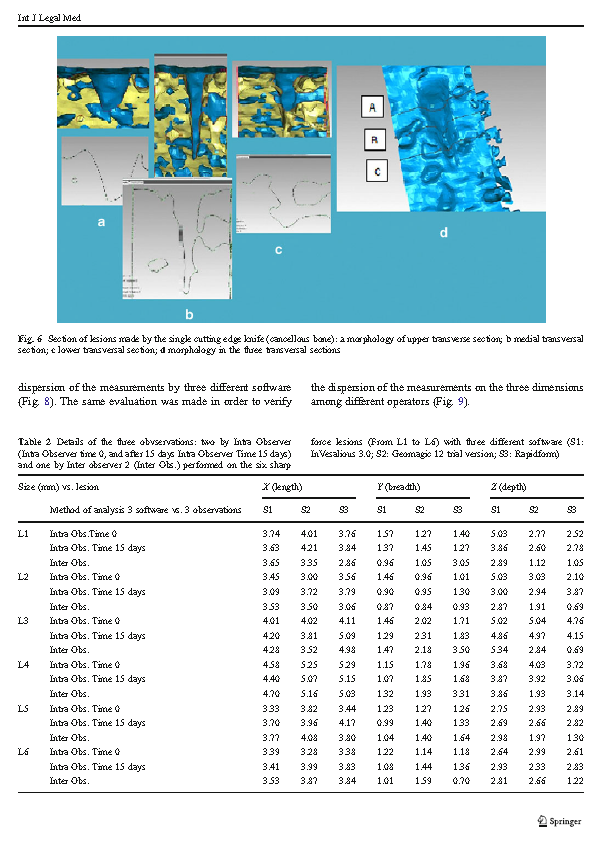
72 71
73 72
74 73
75 74

76 75
77 CAPITOLO 6 Application of high resolution pqct analysis to forensic cases for the assessment of bone trauma: a technical note Rubinacci A, Tresoldi D, Villa I, Rizzo G, Gaudio D, De Angelis D, Gibelli D, Cattaneo C. To be submitted International Journal of Legal Medicine 76
78 Technical note Application of high resolution pqct analysis to forensic cases for the assessment of bone trauma: a technical note Rubinacci A 1,MD, Tresoldi D 2, Villa I 1, PhD, Rizzo G 2, Gaudio D 3,BSc De Angelis D 3 MD. PhD MD, Gibelli D 3 MD PhD Cattaneo C 3, MD, PhD 1 Bone Metabolism Unit Scientific Institute San Raffaele, Milano 2 Institute of Molecular Bioimaging and Physiology, CNR, Segrate (Milan), Italy 3 LABANOF, Laboratorio di Antropologia e Odontologia Forense Sezione di Medicina Legale Dipartimento di Scienze Biomediche per la Salute Università degli Studi di Milano Abstract The evaluation of bone trauma has been considerably improved by the introduction of the most recent technologies; in the last years the peripheral quantitative computed tomography (pqct) has found new fields of application in clinical medicine, but none of them concerns the forensic practice. This study exposes the potential of pqct applied to forensic scenario throughout a real case, concerning a mummified corpse of a 16 year old girl. The autopsy pointed out several sharp force wounds at the chest. Among the others, a specific penetrating lesion affected the body of the first thoracic vertebra, deepening within the underlying cancellous bone. A pqct scanner was used for the measurements (Research SA+; Stratec Medizintechnik GmbH, Pforzheim, Germany). A more precise reconstruction of the path of the lesion within the trabecular bone was reached, with more details concerning the morphological characteristics of the lesion inside the vertebral bone and the elaboration of a 3D model was created, which allowed the operator to define the volume of the lack of tissues related to the lesion. The application of pqct scan proved to be a potentially useful tool for assessment of bone trauma, although further studies are needed in order to verify its reliability. Keywords: forensic anthropology; pqct; bone trauma, sharp force 77
79 Introduction An accurate analysis of bone fractures is crucial for determining cause and manner of death; the accurate fracture interpretation is essential for identifying the location of impact sites, establishing multiple impact sites, sequence of blows and the characteristics of the weapon (1). From this point of view the study of bone lesions is one of the main topic in forensic anthropology and pathology. In particular the analysis of Sharp force wounds, and especially stab wounds, is as crucial as problematic: the correct interpretation of the morphology of lesions on bone tissue may be fundamental to recognize the tool used and reconstructing the way of inflicting trauma, however the small dimensions of lesions often don t allow an exhaustive interpretation about the weapon that produced them. This topic has known in the last years a crucial development by the application of most advanced technologies such as scanning electron microscopy coupled with X-ray energy dispersive spectrometry (SEM-EDS) (2,3) and traditional CT scan and NMR technologies (4,5,6,7), and the application of new techniques which allow the operator to draw a more precise visualization of bone structure, such as cone beam computed tomography (8,9). So what are the radiological technologies more effective to document and investigate very small lesions, but fundamental in order to reconstruct manner of death, like sharp wounds? The Micro-CT technology allows us to reach very high in resolution compared to conventional CT (in general isometric voxel was from 10 to 100 microns, the resolution depends of the type of Micro-CT), however bones may have to undergo partial destruction. Cone Beam technology (used in the odontological field) can also be used on middle or little size samples, without causing their distruction. The first studies concerning the employment of this technology for the documentation of Sharp Wounds on spongy bones seem to lead to promising results (9). The natural history of forensic sciences is characterized by the progressive implementation in the forensic practice of the most advanced technologies, usually previously developed with clinical purposes, with a consequent improvement of morphological and metrical details of bone structures. This has led in some cases to the blind application of such techniques to the forensic cases, without a preliminary evaluation concerning the specific issues which can be adequately and specifically evaluated by the new technologies, usually determined by the idea that technology means more information. This point of view was denied in the last years by different authors which underlined the existence of specific factors of variability which affect the analysis of corpses and define the complementary nature of the new techniques with regards to 78
80 the classical morphological autoptical and forensic anthropological approaches (10). On the other hand, some methods may bring about relevant improvements in the field of bone assessment, since they may meet specific limits which cannot be faced by other addition, the specific properties of the pqct make easier the analysis of the trabecular bone, which is traditionally difficult to assess by the classic radiological methods. methodologies or the traditional At the moment, pqct has been applied to morphological analysis. This is the case of the different field of research, especially for the morphological evaluation of signs of trauma evaluation of osteoporosis and bone on trabecular bone, which is usually difficult to assess by the macroscopic, microscopic distribution, thanks to its accuracy in analysis of trabecular bone (13-14, 15). In addition, (SEM-EDX) and radiological methods recently it has been used for investigating the because of the most irregular disposition of bone trabeculae and the absence of the compact structure of the cortical bone (moreover the traditional casting techniques are problematic on cancellous bone). The recent introduction of Peripheral Quantitive Computerized Tomography (pqct) may give some help in this sensitive field of research: it is a tomographic technique able to assess quantitatively the densitometric properties of bone. In addition, current pqct scanners have a sufficient resolution (70 µm) to anatomy of dental root canals and canal volumes changes after instrumentation (16). However, at the moment no study has been yet still performed concerning the possible application of such technology to the forensic scenario, for example for assessing bone trauma in trabecular bone. This study aims at providing the first example of application of pqct to a real forensic case in order to highlight the advantages which may derive from the use of such modern technique. visualize the trabecular network of human bone in the three spatial dimensions (11), and therefore allow the operator to reconstruct an accurate geometrical and densitometric model of bone which takes into account both macro and microstructural components (12). The use of specific developed software can provide a 3D model of the bone specimen, based on the information obtained by the scanner. In this way, this technology allows the observer to obtain a virtual model of the bone sample which can be analysed in three dimensions; in Case report In 2010 a mummified corpse of a 16 year old girl was found in a church; the subject was seen alive for the last time in The autopsy pointed out several sharp force wounds at the chest. Ribs, cervical and thoracic vertebrae and the scapulae were recovered and cleaned by mummified tissues in order to analyse in depth the bone lesions. Among the others, a specific penetrating lesion affected the body of the first thoracic 79
81 vertebra (Fig. 1), deepening within the underlying cancellous bone. The morphology of the lesion and its position inside a bone rich in trabecular bone required more detailed radiological tests, among which a conventional TC scan which gave general indications concerning the depth and breadth of the lesion. A pqct scanner was therefore used for the measurements (Research SA+; Stratec Medizintechnik GmbH, Pforzheim, Germany). This translation rotation scanner works with a specially developed X-ray tube with a 50-lm spot size (high voltage 50 kv, anode current <0.3 ma, mean X-ray energy 37 kev, energy distribution after filtration 18 kev full width half maximum [FWHM]). The detector-system consists of 12 miniature semiconductor crystals with amplifiers. The precision error supplied by the manufacture for density measurement in vivo is around 1.5%. All images were obtained with 360 projections, with a section thickness of 100 µm and at in-plane pixel size of 100 x 100 µm. The thoracic vertebra was positioned in the centre of a perspex container made for this purpose and total scanning time was 4 hours. A more precise reconstruction of the path of the lesion within the trabecular bone was reached (Fig. 2), with more details concerning the morphological characteristics of the lesion inside the vertebral bone. In addition, a 3D reconstruction and visualization of the structure of interest was created. Briefly the procedure consisted in the following steps: 1) slice by slice bone contour extraction using an already validated algorithm to divide bone tissue from background [13, 14]; 2) definition of the area characterized by lack of tissues due to the lesion and its contour; 3) starting from these contours (bone and lesion area), generation of a 3D model as a polygonal mesh structure; 4) 3D rendering of the mesh model, characterizing, each structure of interest with specific colour, brightness and transparency (Fig. 3). In this way, the visualization of the inner walls of the lesion was also possible with the chance of taking measurements of the linear and volume parameters (Fig. 4). Discussion and conclusion. In the last year the forensic practice has seen a constant and progressive increase of application of the most modern technologies, usually implemented from the clinical context. The recent introduction of pqct has considerably increased the precision of analysis of the trabecular bone, which is usually difficult to assess by traditional microscopic or radiological techniques. From this point of view the present case study has shown an increased definition of one lesions within the spongeous bone; in addition, the details of the 3D models provide an improved evaluation of the morphology and metrical parameters of bone lesions, with respect to the conventional CT scan which fails in defining the lesions edges within the trabecular bone. In addition, the 3D 80
82 reconstruction of the volume showing lack of tissue allows the operator to extrapolate relevant information concerning the metrical parameters of the lesion, with a clear amelioration for what concerns the reconstruction of the used weapon. Finally, the pqct technology allows to obtain a good resolution (70 µm, more high of CT Cone Beam) This first example of the application of such technology to a forensic case shows the potential of pqct for the assessment of bone trauma, although, as observed for other modern technologies applied to the forensic field, some points of criticism need to be overcome, especially for what concerns the correct evaluation of the lesion path within the trabecular bone. Further studies are therefore needed in order to correctly define the limits of such technology. 81
 with details of the same lesion by")

83 Fig. 1: detail of the sharp force lesion on the first thoracic vertebra (in the red square) with details of the same lesion by stereomicroscopy Fig. 2: visualization of the vertebral lesion by use of pqct; in the red square, image of the same lesion by conventional CT scan 82
84 Fig. 3: rapresentative 3D rendering of the model of the first thoracic vertebra acquired by pqct; the bone is coloured in yellow and shown in transparency, the bone lesion is in red. Fig. 4: extrapolation of the volume and inner morphology of the bone lesion by the 3D model acquired by the pqct 83
85 References 1) Burd DQ, Gilmore AE, Individual and class characteristics of tools, J Forensic Sci 2001;46(2): ) Pechnikovà M, Porta D, Mazzarelli D, Rizzi A, Drozdova E, Gibelli D, Cattaneo C, Detection of metal residues on bone using SEM-EDS. Part I: blunt force injury, Forensic Science International, 2012, in press 3) Gibelli D, Mazzarelli D, Porta D, Rizzi A, Cattaneo C, Detection of metal residues on bone using SEM- EDS Part II: sharp force injury, Forensic Sci Int 2012, in press 4) Buck U, Christe A, Naether S, Ross S, Thali MJ, Virtopsy - noninvasive detection of occult bone lesions in postmortem MRI: additional information for traffic accident reconstruction. Int J Legal Med. 2009;123(3): ) Thali MJ, Taubenreuther U, Karolczak M, Braun M, Brueschweiler W, Kalender WA, Dirnhofer R, Forensic microradiology: micro-computed tomography (Micro-CT) and analysis of patterned injuries inside of bone, J Forensic Sci. 2003;48(6): ) Capuani C et al (2011) Deciphering the elusive nature of sharp bone trauma using epifluorescence macroscopy: a comparison study multiplexing classical imaging approaches, Int J Legal Med [Epub ahead of print]. 7) Pounder DJ et al (2011) Virtual Casting of Stab Wounds in Cartilage Using Micro-Computed Tomography. Am J Forensic Med Pathol 32(2): ) Von See C, Bormann KH, Schumann P, Goetz F, Gellrich NC, Rücker M, Forensic imaging of projectiles using cone-beam computed tomography, Forensic Sci Int. 2009;190(1-3): ) Gaudio D, Di Giancamillo M, Gibelli D, Galassi A, Cerutti E, Cattaneo C., Does cone beam CT actually ameliorate stab wound analysis in bone? International Journal of Legal Medicine (2013) (In press). 10) Cattaneo C, Marinelli E, Di Giancamillo A, Di Giancamillo M, Travetti O, Vigano' L, Poppa P, Porta D, Gentilomo A, Grandi M, Sensitivity of autopsy and radiological examination in detecting bone fractures in an animal model: implications for the assessment of fatal child physical abuse. Forensic Sci Int. 2006;164(2-3): ) MacNeil JA, Boyd SK, Accuracy of high-resolution peripheral quantitative 84
86 computed tomography for measurement of bone quality, Med Eng Phys 2007(29): ) Boutroy S, Bouxsein ML, Munoz F, Delmas PD, In vivo assessment of trabecular bone microarchitecture by high-resolution peripheral quantitative computed tomography, J Clin Endocrinol Metab 2005(90): ) Rizzo G, Tresoldi D, Scalco E, Mendez M, Bianchi AM, Moro GL, Rubinacci A, Automatic segmentation of cortical and trabecular components of bone specimens acquired by pqct, 30 th Annual Internatonal IEEE EMBS Conference, Vancouver, British Columbia, Canada, August ) Rizzo, G, Scalco E, Tresoldi D, Villa I, Moro GL, Lafortuna CL, Rubinacci A, An automatic segmentation method for regional analysis of femoral neck images acquired by pqct, Ann Biomed Eng 2011;39(1): ) Rubinacci A, Tresoldi D, Scalco E, Villa I, Adorni F, Moro GL, Fraschini GF, Rizzo G, Comparative highresolution pqct analysis of femoral neck indicates different bone mass distribution in osteoporosis and osteoarthritis, Osteoporos Int 2012:23: ) Sberna MT, Rizzo G, Zacchi E, Capparè P, Rubinacci A, A preliminary study of the use of peripheral quantitative computed tomography for investigating root canal anatomy, Int J Endodontic J 2009;42:
87 CAPITOLO 7 The application of cone-beam CT in the aging of bone calluses: a new perspective? Cappella A, Amadasi A, Gaudio D, Gibelli D, Borgonovo M, Di Giancamillo M, Cattaneo C. International journal of legal medicine (2013) 127:
88 87
89 88
90 89
91 90
92 91

93 92
Le strumentazioni laser scanning oriented per i processi di censimento anagrafico dei patrimoni
 CONVEGNO FACILITY MANAGEMENT: LA GESTIONE INTEGRATA DEI PATRIMONI PUBBLICI GENOVA FACOLTA DI ARCHITETTURA 06.07.2010 Le strumentazioni laser scanning oriented per i processi di censimento anagrafico dei
Elementi di Psicometria con Laboratorio di SPSS 1
 Elementi di Psicometria con Laboratorio di SPSS 1 29-Analisi della potenza statistica vers. 1.0 (12 dicembre 2014) Germano Rossi 1 germano.rossi@unimib.it 1 Dipartimento di Psicologia, Università di Milano-Bicocca
La codifica delle immagini
 La codifica delle immagini La digitalizzazione La digitalizzazione di oggetti legati a fenomeni di tipo analogico, avviene attraverso due parametri fondamentali: 1. Il numero dei campionamenti o di misurazioni
IL RILIEVO TOPOGRAFICO CON LASER SCANNER
 IL RILIEVO TOPOGRAFICO CON LASER SCANNER fb.m. s.r.l. 1 Il continuo sviluppo della tecnica di rilievo e la ricerca continua del miglior risultato ore-lavoro, ha prodotto la strumentazione per il rilievo
Estendere Lean e Operational Excellence a tutta la Supply Chain
 Estendere Lean e Operational Excellence a tutta la Supply Chain Prof. Alberto Portioli Staudacher www.lean-excellence.it Dipartimento Ing. Gestionale Politecnico di Milano alberto.portioli@polimi.it Lean
3DE Modeling Professional
 3DE Modeling Professional 3DE Modeling Professional è la parte di 3DE Modeling Suite che si occupa della modellazione 3D automatica di oggetti ed edifici a partire da nuvole di punti ottenute con scanner
IsoStereo 3D: il sistema generale ed automatizzato per ricostruzione tridimensionale
 IsoStereo 3D: il sistema generale ed automatizzato per ricostruzione tridimensionale Isomorph srl, Giugno 2008 Caratteristiche del programma: IsoStereo 3D elabora immagini stereoscopiche e ricostruisce
RILIEVO LASER SCANNER PARETE MARMOREA XXXXXXXXXXXXX
 RILIEVO LASER SCANNER PARETE MARMOREA XXXXXXXXXXXXXXXXXX PROVA n. 3651/XX Committente: Relatore: XXXXXXXXXXXXX ing. Georg Schiner Vista della parete di intarsio marmoreo XX/88/09/mt Bolzano, 9 Settembre
Archivi e database. Prof. Michele Batocchi A.S. 2013/2014
 Archivi e database Prof. Michele Batocchi A.S. 2013/2014 Introduzione L esigenza di archiviare (conservare documenti, immagini, ricordi, ecc.) è un attività senza tempo che è insita nell animo umano Primi
Il gis nelle indagini del territorio: Ricostruzione tridimensionale del sottosuolo
 Il gis nelle indagini del territorio: Ricostruzione tridimensionale del sottosuolo A. Genito, L. Donate Blázquez, U. Malvone, Strago S.p.a. andrea.genito@strago.it laura.donate@strago.it ubaldo.malvone@strago.it
UNIVERSITÀ DEGLI STUDI DI MILANO FACOLTÀ DI STUDI UMANISTICI Corso di laurea triennale in Scienze umanistiche per la comunicazione
 UNIVERSITÀ DEGLI STUDI DI MILANO FACOLTÀ DI STUDI UMANISTICI Corso di laurea triennale in Scienze umanistiche per la comunicazione LA RETE SOCIALE PER COMUNICARE L'AMBIENTE: SOCIAL NETWORK ED ECOLOGIA
UNIVERSITÀ DEGLI STUDI GUGLIELMO MARCONI
 UNIVERSITÀ DEGLI STUDI GUGLIELMO MARCONI FACOLTÀ DI SCIENZE E TECNOLOGIE APPLICATE CORSO DI LAUREA IN SCIENZE GEO-CARTOGRAFICHE, ESTIMATIVE ED EDILIZIE TECNICHE DI MODELLAZIONE 3D A PARTIRE DA DATI RILEVATI
Novità 2013 RILIEVO 3D CON LASER SCANNER
 Novità 2013 RILIEVO 3D CON LASER SCANNER Il rilievo con Laser Scanner produce una nuvola di punti che riproduce fedelmente l intera area interessata dall intervento. Che si tratti del rilievo di un edificio,
LA DIAGNOSTICA MOLECOLARE E I TUMORI DEL SANGUE
 LA DIAGNOSTICA MOLECOLARE E I TUMORI DEL SANGUE UNA NUOVA FRONTIERA NELLA DIAGNOSI DI ALCUNI TUMORI La diagnostica molecolare ha l obiettivo di accertare un ampia varietà di patologie (infettive, oncologiche
3DEverywhere S.r.l. sito web: http://www.3deverywhere.com
 3DE Modeling Analyzer 3DE Modeling Analyzer permette di effettuare un'analisi accurata dei modelli ottenuti con 3DE Modeling Professional. 3DE Modeling Analyzer consente: il caricamento di singole viste
Guida Compilazione Piani di Studio on-line
 Guida Compilazione Piani di Studio on-line SIA (Sistemi Informativi d Ateneo) Visualizzazione e presentazione piani di studio ordinamento 509 e 270 Università della Calabria (Unità organizzativa complessa-
CS 7400. La soluzione ideale per passare al digitale
 CS 7400 La soluzione ideale per passare al digitale Immagini più grandi e di alta qualità Una scelta digitale conveniente ed economicamente vantaggiosa Mentre molti sistemi di radiografia digitale possono
SCAN & GO IL NUOVO MODO DI FARE TOPOGRAFIA
 SCAN & GO IL NUOVO MODO DI FARE TOPOGRAFIA Scan&Go è una nuova metodologia per il rilievo topografico del territorio che utilizza strumentazione laser scanner abbinata a ricevitori GPS. Il sistema, installato
Sistema di ripresa con fotocamera digitale fissa Calibrazione
 Sistema di ripresa con fotocamera digitale fissa Calibrazione TUTORIAL CALIBRAZIONE FOTOCAMERA Sommario 1. Interfacciamento PC... 3 2. Collocamento fotocamera... 4 3. Distanza di ripresa... 5 4. Interfacciamento
Corso di Informatica
 CdLS in Odontoiatria e Protesi Dentarie Corso di Informatica Prof. Crescenzio Gallo crescenzio.gallo@unifg.it Immagini radiologiche!2 PACS PACS è l'acronimo anglosassone di Picture archiving and communication
Progetto IDENTITAS: Formazione agli operatori di Bilancio di Competenze
 Progetto IDENTITAS: Formazione agli operatori di Bilancio di Competenze Provincia di Roma Anno 2005 Indice Il problema affrontato...3 Obiettivi attesi/risultati raggiunti...3 Soggetti coinvolti...3 Il
"Rapid Prototyping: studio delle possibili sorgenti di errore"
 "Rapid Prototyping: studio delle possibili sorgenti di errore" Facoltà di Ingegneria Civile ed Industriale Corso di laurea in Ingegneria Meccanica Laureando: Daniele Cortis Relatore: Ing. Francesca Campana
Il campionamento. La digitalizzazione. Teoria e pratica. La rappresentazione digitale delle immagini. La rappresentazione digitale delle immagini
 ACQUISIZIONE ED ELABORAZIONE DELLE IMMAGINI Teoria e pratica La digitalizzazione La digitalizzazione di oggetti legati a fenomeni di tipo analogico, avviene attraverso due parametri fondamentali: Il numero
IL CICLO DI VITA DEL PROGETTO. Elementi essenziali di progetto. Fasi e tappe Gli Approcci
 UNIVERSITA MILANO BICOCCA Corso di laurea di primo livello in servizio sociale anno accademico 2009-2010 Progettare il sociale Prof. Dario A. Colombo IL CICLO DI VITA DEL PROGETTO Elementi essenziali di
UNIVERSITA DEGLI STUDI ROMA TRE
 UNIVERSITA DEGLI STUDI ROMA TRE COMUNITA DI PRATICA E COMUNICAZIONE ON LINE : IL CASO MASTER GESCOM RELATORE Prof.ssa Giuditta ALESSANDRINI Laureanda Chiara LOLLI Matr. n. 203597 CORRELATORE Prof.ssa Isabella
Dispensa di Informatica I.1
 IL COMPUTER: CONCETTI GENERALI Il Computer (o elaboratore) è un insieme di dispositivi di diversa natura in grado di acquisire dall'esterno dati e algoritmi e produrre in uscita i risultati dell'elaborazione.
Capitolo V : Il colore nelle immagini digitali
 Capitolo V : Il colore nelle immagini digitali Lavorare con il colore nelle immagini digitali L uso dei colori nella visione computerizzata e nella computer grafica implica l incorrere in determinate problematiche
Sistema di diagnosi CAR TEST
 Data: 30/09/09 1 di 7 Sistema di diagnosi CAR TEST Il sistema di diagnosi CAR TEST venne convenientemente utilizzato per: - verificare che la scocca di un veicolo sia dimensionalmente conforme ai disegni
creazione di oggetti
 La stampa 3D (conosciuta nel mondo anche nella versione inglese del termine come 3D printing) è una nuova tecnologia sviluppata a negli ultimi anni che costituisce una vera e propria evoluzione della stampa
Misure senza contatto
 Misure senza contatto Remo Sala - remo.sala@polimi.it VBLab Dipartimento di Meccanica Politecnico di Milano 03 luglio 2009 - Corso presso medicina legale 1 Messaggio: posso creare delle copie 2 Messaggio:
SCAN&GO IL NUOVO MODO DI FARE TOPOGRAFIA. www.scan-go.it www.scan-go.eu V.03.1213
 SCAN&GO IL NUOVO MODO DI FARE TOPOGRAFIA V.03.1213 www.scan-go.it www.scan-go.eu SCAN&GO SYSTEM IL NUOVO MODO DI FARE TOPOGRAFIA Scan&Go è una nuova metodologia per il rilievo topografico del territorio
Sicurezza Aziendale: gestione del rischio IT (Penetration Test )
") Sicurezza Aziendale: gestione del rischio IT (Penetration Test ) Uno dei maggiori rischi aziendali è oggi relativo a tutto ciò che concerne l Information Technology (IT). Solo negli ultimi anni si è iniziato
Sicurezza Aziendale: gestione del rischio IT (Penetration Test ) Uno dei maggiori rischi aziendali è oggi relativo a tutto ciò che concerne l Information Technology (IT). Solo negli ultimi anni si è iniziato
Data Alignment and (Geo)Referencing (sometimes Registration process)
Referencing (sometimes Registration process)") Data Alignment and (Geo)Referencing (sometimes Registration process) All data aquired from a scan position are refered to an intrinsic reference system (even if more than one scan has been performed) Data
Data Alignment and (Geo)Referencing (sometimes Registration process) All data aquired from a scan position are refered to an intrinsic reference system (even if more than one scan has been performed) Data
Più che una protesi su impianto. Un sorriso naturale.
 Informazioni per i pazienti: protesi fisse su impianti Più che una protesi su impianto. Un sorriso naturale. Più che funzionalità. Una ritrovata sicurezza. Soddisfatti della protesi tradizionale? Piccoli
IMMAGINE BITMAP E VETTORIALI
 BITMAP VETTORIALE VETTORIALE BITMAP IMMAGINE BITMAP è una immagine costituita da una matrice di Pixel. PIXEL (picture element) indica ciascuno degli elementi puntiformi che compongono la rappresentazione
SOFTWARE PER LA RILEVAZIONE DEI TEMPI PER CENTRI DI COSTO
 SOFTWARE PER LA RILEVAZIONE DEI TEMPI PER CENTRI DI COSTO Descrizione Nell ambito della rilevazione dei costi, Solari con l ambiente Start propone Time&Cost, una applicazione che contribuisce a fornire
www.andreatorinesi.it
 La lunghezza focale Lunghezza focale Si definisce lunghezza focale la distanza tra il centro ottico dell'obiettivo (a infinito ) e il piano su cui si forma l'immagine (nel caso del digitale, il sensore).
API e socket per lo sviluppo di applicazioni Web Based
 API e socket per lo sviluppo di applicazioni Web Based Cosa sono le API? Consideriamo il problema di un programmatore che voglia sviluppare un applicativo che faccia uso dei servizi messi a disposizione
DIFFERENZIARE LE CAMPAGNE DI MARKETING La scelta del canale adeguato
 Via Durini, 23-20122 Milano (MI) Tel.+39.02.77.88.931 Fax +39.02.76.31.33.84 Piazza Marconi,15-00144 Roma Tel.+39.06.32.80.37.33 Fax +39.06.32.80.36.00 www.valuelab.it valuelab@valuelab.it DIFFERENZIARE
Rilevazione tramite Photoscan! Esempio applicativo
 Rilevazione tramite Photoscan Esempio applicativo Considerazioni generali Un quadro completo dell'elemento da rilevare con eventuali salti di quota, pendenze, punti di inaccessibilità, vegetazione ecc.
BODY SCANFIT : il primo body scanner 3D portatile per digitalizzare in 4 secondi il mondo reale
 BODY SCANFIT : il primo body scanner 3D portatile per digitalizzare in 4 secondi il mondo reale Professionisti della moda e del su misura, specialisti di medicina, di ergonomia, ma anche operatori cinema
Ottica fotografica. Lezioni per il corso di Fisica per gli studenti del diploma di Ottica
 Ottica fotografica Lezioni per il corso di Fisica per gli studenti del diploma di Ottica Alessandro Farini alessandro.farini@inoa.it Istituto Nazionale di Ottica Applicata-CNR 2 luglio 2009 Alessandro
63 7. Quale geometria per la computer grafica? 75 8. L omografia e l affinità nella digitalizzazione e georeferenziazione
 Indice 7 Presentazione 9 Premessa 11 Introduzione 13 1. Rilevamento ed oggetto 19 2. La stazione totale 23 3. La procedura generale 33 4. Dai punti al modello tridimensionale 45 5. Il modello tridimensionale
SymCAD/C.A.T.S. modulo Canali Schema
 SymCAD/C.A.T.S. modulo Canali Schema Il modulo Ventilazione Standard permette di effettuare la progettazione integrata (disegno e calcoli) in AutoCAD di reti di canali aria (mandata e ripresa). Il disegno
Rilievo morfologico del manto stradale
 Rilievo morfologico del manto stradale Federico Ferrari L utilizzo del Laser Scanner D Cyrax System nel campo dell incidentistica stradale per perizie legali è collocabile all interno dei programmi di
Il rilievo 3D dei piccoli oggetti: strumenti, metodi e applicazioni
 Nikon Instruments s.p.a Microgeo s.r.l La memoria geometrica degli oggetti: dal millimetro al micron 20 Maggio 2014 Il rilievo 3D dei piccoli oggetti: strumenti, metodi e applicazioni Dott. e Arch. Sergio
Imprenditorialità e Innovazione per l Internazionalizzazione delle PMI
 Corso di Alta Formazione Imprenditorialità e Innovazione per l Internazionalizzazione delle PMI Evento Finale 25 marzo 2013 -Bergamo Palazzo dei Contratti e delle Manifestazioni BERGAMO SVILUPPO AZIENDA
ALLEGATO B. Nel corso degli anni il monitoraggio ha previsto nelle diverse annualità:
 ALLEGATO B Il modello di monitoraggio Il gruppo di Progetto di PQM, nell anno scolastico 2010-2011 ha costituito un gruppo di lavoro tecnico composto dal gruppo di progetto stesso e dai ricercatori dei
COME VIENE REALIZZATA UNA RICERCA SPERIMENTALE IN BIOLOGIA MOLECOLARE?
 COME VIENE REALIZZATA UNA RICERCA SPERIMENTALE IN BIOLOGIA MOLECOLARE? A Flusso di attività B - INPUT C Descrizione dell attività D RISULTATO E - SISTEMA PROFESSIONALE 0. RICHIESTA DI STUDIARE E/O INDIVIDUARE
itime Chiaramente inclusa la stampa del cartellino presenze come previsto dalle normative
 itime itime Il software di rilevazione presenze itime rappresenta lo strumento ideale per l automatizzazione della gestione del personale. L ampia presenza dei parametri facilita l operatore nel controllo
PointCloud 7.0 Anno 2011
 PointCloud 7.0 Anno 2011 Compatibilità con AutoCAD 2007-2012 ed AutoCAD 2007-2012 Supporto del formato nativo delle nuvole di punti di AutoCAD (PCG) e miglioramento delle performance riguardo la velocità
Università degli Studi di Napoli Federico II Facoltà di Ingegneria. Corso di. Sistemi Distribuiti. Prof. Stefano Russo. Field Failure Data Analysis
 Università degli Studi di Napoli Federico II Facoltà di Ingegneria Corso di Sistemi Distribuiti Prof. Stefano Russo Domenico Cotroneo Failure Data Analysis (FDA) (1/2) I mezzi per la dependability possono
L evoluzione del mosaico digitale
 L evoluzione del mosaico digitale MOSAIC PROJECT SUPPORTA L INTERO PROCESSO DEL MOSAICO DALLA FASE CREATIVA A QUELLA PRODUTTIVA, SIA PER QUANTO RIGUARDA L INDUSTRIALE E L ARTISTICO. LE CARATTERISTICHE
Introduzione all analisi dei segnali digitali.
 Introduzione all analisi dei segnali digitali. Lezioni per il corso di Laboratorio di Fisica IV Isidoro Ferrante A.A. 2001/2002 1 Segnali analogici Si dice segnale la variazione di una qualsiasi grandezza
Sinossi dei disegni di studio
 Sinossi dei disegni di studio di intervento osservazionali serie di casi trasversale ecologici individuali quasisperimentali sperimentali (RCT) coorte caso-controllo Studi caso-controllo Il punto di partenza
MODULO 5 Appunti ACCESS - Basi di dati
 MODULO 5 Appunti ACCESS - Basi di dati Lezione 1 www.mondopcnet.com Modulo 5 basi di dati Richiede che il candidato dimostri di possedere la conoscenza relativa ad alcuni concetti fondamentali sui database.
Martina Agazzi. Corso di Tecniche plastiche contemporanee. Prof. Carlo Lanzi
 Martina Agazzi Corso di Tecniche plastiche contemporanee Prof. Carlo Lanzi RILEVAMENTO 3D DI UN OGGETTO ARTISTICO (SCULTURA) Luce strutturata Dispositivo portatile, facilmente trasportabile digitalizzazione
La radiochirurgia tramite Gamma Knife Informazioni per i pazienti
 La radiochirurgia tramite Gamma Knife Informazioni per i pazienti La radiochirurgia tramite Gamma Knife è un metodo ampiamente diffuso per il trattamento di un area selezionata del cervello, detta bersaglio.
Excel. A cura di Luigi Labonia. e-mail: luigi.lab@libero.it
 Excel A cura di Luigi Labonia e-mail: luigi.lab@libero.it Introduzione Un foglio elettronico è un applicazione comunemente usata per bilanci, previsioni ed altri compiti tipici del campo amministrativo
Laboratorio di Scienze Forensi. Pin s.c.r.l. University of Florence
 Laboratorio di Scienze Forensi Pin s.c.r.l. University of Florence Chi siamo Il FORLAB è nato nel 2010 nell Università di Firenze dall incontro di professionisti del settore forense e ricercatori accademici
-Rilievo diretto Laboratori -Rilievo Aula 9 edificio A. Alessio Tirapelle Mirko Mondini Daniel Colombelli Irene Gregori
 -Rilievo diretto Laboratori -Rilievo Aula 9 edificio A Alessio Tirapelle Mirko Mondini Daniel Colombelli Irene Gregori Operazioni di misura a diretto contatto con l oggetto da rilevare; è possibile il
Automazione Industriale (scheduling+mms) scheduling+mms. adacher@dia.uniroma3.it
 scheduling+mms. adacher@dia.uniroma3.it") Automazione Industriale (scheduling+mms) scheduling+mms adacher@dia.uniroma3.it Introduzione Sistemi e Modelli Lo studio e l analisi di sistemi tramite una rappresentazione astratta o una sua formalizzazione
Automazione Industriale (scheduling+mms) scheduling+mms adacher@dia.uniroma3.it Introduzione Sistemi e Modelli Lo studio e l analisi di sistemi tramite una rappresentazione astratta o una sua formalizzazione
L innovativa tecnologia digitale per realizzare splendidi sorrisi
 L innovativa tecnologia digitale per realizzare splendidi sorrisi Universo NemoBridge è l unico sistema esclusivo di tecnologia avanzata capace di offrire una soluzione completa per l implantologia. Un
gestione e modifica di immagini fotografiche digitali
 gestione e modifica di immagini fotografiche digitali il colore e le immagini la gestione delle immagini Il computer è in grado di gestire le immagini in formato digitale. Gestire vuol dire acquisirle,
La riforma del servizio di distribuzione del
 CReSV Via Röntgen, 1 Centro Ricerche su Sostenibilità e Valore 20136 Milano tel +39 025836.3626 La riforma del servizio di distribuzione del 2013 gas naturale In collaborazione con ASSOGAS Gli ambiti territoriali
Il database management system Access
 Il database management system Access Corso di autoistruzione http://www.manualipc.it/manuali/ corso/manuali.php? idcap=00&idman=17&size=12&sid= INTRODUZIONE Il concetto di base di dati, database o archivio
GUIDA AL CALCOLO DEI COSTI DELLE ATTIVITA DI RICERCA DOCUMENTALE
 GUIDA AL CALCOLO DEI COSTI DELLE ATTIVITA DI RICERCA DOCUMENTALE L applicazione elaborata da Nordest Informatica e disponibile all interno del sito è finalizzata a fornirvi un ipotesi dell impatto economico
Introduzione. Classificazione di Flynn... 2 Macchine a pipeline... 3 Macchine vettoriali e Array Processor... 4 Macchine MIMD... 6
 Appunti di Calcolatori Elettronici Esecuzione di istruzioni in parallelo Introduzione... 1 Classificazione di Flynn... 2 Macchine a pipeline... 3 Macchine vettoriali e Array Processor... 4 Macchine MIMD...
Il controllo dei rischi operativi in concreto: profili di criticità e relazione con gli altri rischi aziendali
 La gestione dei rischi operativi e degli altri rischi Il controllo dei rischi operativi in concreto: profili di criticità e relazione con gli altri rischi aziendali Mario Seghelini 26 giugno 2012 - Milano
QUALITÀ E SICUREZZA PER I NOSTRI PAZIENTI
 QUALITÀ E SICUREZZA PER I NOSTRI PAZIENTI L ACCREDITAMENTO INTERNAZIONALE ALL ECCELLENZA Fondazione Poliambulanza ha ricevuto nel dicembre 2013 l accreditamento internazionale all eccellenza da parte di
Librerie digitali. Video. Gestione di video. Caratteristiche dei video. Video. Metadati associati ai video. Metadati associati ai video
 Video Librerie digitali Gestione di video Ogni filmato è composto da più parti Video Audio Gestito come visto in precedenza Trascrizione del testo, identificazione di informazioni di interesse Testo Utile
Comune di San Martino Buon Albergo
 Comune di San Martino Buon Albergo Provincia di Verona - C.A.P. 37036 SISTEMA DI VALUTAZIONE DELLE POSIZIONI DIRIGENZIALI Approvato dalla Giunta Comunale il 31.07.2012 INDICE PREMESSA A) LA VALUTAZIONE
CONVENZIONE UNIVERSITÀ DI PERUGIA DELTATECH. Rapporto Attività di Ricerca. Prove ad impatto su laminati compositi con.
 CONVENZIONE UNIVERSITÀ DI PERUGIA DELTATECH Rapporto Attività di Ricerca Prove ad impatto su laminati compositi con Ball Drop Tester Prof. L. Torre Fase 1: Terni 18/6/214 METODI Sono stati effettuati test
La libertà di sorridere
 Impianti da vivere just smile La libertà di sorridere Il sorriso apre le porte, dà fiducia, a chi lo fa e a chi lo riceve, migliora le relazioni. Una dentatura sana è sinonimo di positività, di vitalità,
3D e Realtà Virtuale
 3D e Realtà Virtuale Modello 3D La costruzione di un modello 3D, così come la realizzazione di un plastico tradizionale, necessita di grande precisione e attenzione conoscitiva. Tale modello convoglia
L ATI composta da Associazione Nuovi Lavori e BIC Puglia s.c.a.r.l., nell ambito del progetto URLO Una rete per le opportunità, PROMUOVE
 L ATI composta da Associazione Nuovi Lavori e BIC Puglia s.c.a.r.l., nell ambito del progetto URLO Una rete per le opportunità, PROMUOVE un PROTOCOLLO D INTESA tra CONSIGLIERA PARITÀ PROVINCIALE DONNE
3DE Modeling Color. E il modulo che si occupa della costruzione di modelli 3D con tessitura a colori.
 3DE Modeling Color E il modulo che si occupa della costruzione di modelli 3D con tessitura a colori. E spesso necessario che alle informazioni geometriche di forma siano abbinate informazioni di colore
GUIDA PER IL DOCENTE ALL UTILIZZO DELL APPLICATIVO ONLINE E PORTFOLIO
 GUIDA PER IL DOCENTE ALL UTILIZZO DELL APPLICATIVO ONLINE E PORTFOLIO http://eportfolio.tqmproject.eu Progetto "TQM Agreement n 2011 1 IT1 LEO05 01873; CUP G72F11000050006 1 SOMMARIO PREMESSA... 3 PAGINA
Guida alla compilazione on-line delle domande di Dote Scuola A.S. 2013-2014 - per le Famiglie INDICE
 Guida alla compilazione on-line delle domande di Dote Scuola A.S. 2013-2014 - per le Famiglie INDICE Introduzione... 2 Riconoscimento del soggetto richiedente da parte del sistema... 2 Elenco dei servizi
TachyCAD 8.0 Anno 2010
 TachyCAD 8.0 Anno 2010 TachyCAD 8.0 contiene molte nuove novità! Le più importanti sono: Nuova struttura del software: nuove soluzioni verticali al posto dell estensione modulare Compatibilità con AutoCAD
LA PROGETTAZIONE DI UN NUOVO STRUMENTO PER IL WEB
 UNIVERSITÀ DEGLI STUDI DI PADOVA FACOLTÀ DI LETTERE E FILOSOFIA CORSO DI LAUREA MAGISTRALE IN STRATEGIE DI COMUNICAZIONE LA PROGETTAZIONE DI UN NUOVO STRUMENTO PER IL WEB LA PROPOSTA DI UN MODELLO MIRATO
Un prototipo di 3D scanner
 Un prototipo di 3D scanner Visual Computing Group 1999 Visual Computing Group 1 Obiettivi Progettazione e realizzazione di uno 3d scanner a basso costo, a partire da hardware comune: una foto camera /
2.0 Gli archivi. 2.1 Inserire gli archivi. 2.2 Archivio Clienti, Fornitori, Materiali, Noleggi ed Altri Costi. Impresa Edile Guida all uso
 2.0 Gli archivi All interno della sezione archivi sono inserite le anagrafiche. In pratica si stratta di tutti quei dati che ricorreranno costantemente all interno dei documenti. 2.1 Inserire gli archivi
COMUNE DI PERUGIA AREA DEL PERSONALE DEL COMPARTO DELLE POSIZIONI ORGANIZZATIVE E DELLE ALTE PROFESSIONALITA
 COMUNE DI PERUGIA AREA DEL PERSONALE DEL COMPARTO DELLE POSIZIONI ORGANIZZATIVE E DELLE ALTE PROFESSIONALITA METODOLOGIA DI VALUTAZIONE DELLA PERFORMANCE Approvato con atto G.C. n. 492 del 07.12.2011 1
GUIDA RAPIDA PER LA COMPILAZIONE DELLA SCHEDA CCNL GUIDA RAPIDA PER LA COMPILAZIONE DELLA SCHEDA CCNL
 GUIDA RAPIDA BOZZA 23/07/2008 INDICE 1. PERCHÉ UNA NUOVA VERSIONE DEI MODULI DI RACCOLTA DATI... 3 2. INDICAZIONI GENERALI... 4 2.1. Non modificare la struttura dei fogli di lavoro... 4 2.2. Cosa significano
Capitolo 3. L applicazione Java Diagrammi ER. 3.1 La finestra iniziale, il menu e la barra pulsanti
 Capitolo 3 L applicazione Java Diagrammi ER Dopo le fasi di analisi, progettazione ed implementazione il software è stato compilato ed ora è pronto all uso; in questo capitolo mostreremo passo passo tutta
RILIEVO CON TECNOLOGIA LASER SCANNER
 RILIEVO CON TECNOLOGIA LASER SCANNER LA SCANSIONE LASER 3D CONSENTE DI PENSARE IN UN MODO NUOVO ALLE ATTIVITA DI ACQUISIZIONE, ANALISI ED ARCHIVIAZIONE DATI, INDIPENDENTEMENTE DAL SETTORE APPLICATIVO IN
ISTITUTO PROFESSIONALE
 Via Resistenza,800 41058 Vignola (Modena) tel. 059 771195 fax. 059 764354 e-mail: mois00200c@istruzione.it www.istitutolevi.it ISTITUTO PROFESSIONALE MANUTENZIONE E ASSISTENZA TECNICA (con qualifica di
lo 2 2-1 - PERSONALIZZARE LA FINESTRA DI WORD 2000
 Capittol lo 2 Visualizzazione 2-1 - PERSONALIZZARE LA FINESTRA DI WORD 2000 Nel primo capitolo sono state analizzate le diverse componenti della finestra di Word 2000: barra del titolo, barra dei menu,
Progetto TIC (trasparenza- informatica-comunicazione)
") Progetto TIC (trasparenza- informatica-comunicazione) Il quadro normativo di seguito riportato evidenzia il ruolo che la Provincia avrà quale ente con funzioni di area vasta che potrà essere di supporto
Progetto TIC (trasparenza- informatica-comunicazione) Il quadro normativo di seguito riportato evidenzia il ruolo che la Provincia avrà quale ente con funzioni di area vasta che potrà essere di supporto
Software di sistema e software applicativo. I programmi che fanno funzionare il computer e quelli che gli permettono di svolgere attività specifiche
 Software di sistema e software applicativo I programmi che fanno funzionare il computer e quelli che gli permettono di svolgere attività specifiche Software soft ware soffice componente è la parte logica
L AP come elemento qualificante della mission didattica degli Atenei
 L AP come elemento qualificante della mission didattica degli Atenei Convegno Nazionale RUIAP Riconoscimento degli apprendimenti e certificazione delle competenze: sfide per l Università e per il Paese
Hub-PA Versione 1.0.6 Manuale utente
 Hub-PA Versione 1.0.6 Manuale utente (Giugno 2014) Hub-PA è la porta d ingresso al servizio di fatturazione elettronica verso la Pubblica Amministrazione (PA) a disposizione di ogni fornitore. Questo manuale
Le immagini digitali. Le immagini digitali. Caterina Balletti. Caterina Balletti. Immagini grafiche. Trattamento di immagini digitali.
 1 Le immagini digitali Le immagini digitali Università IUAV di venezia Trattamento di immagini digitali immagini grafiche immagini raster immagini vettoriali acquisizione trattamento geometrico trattamento
SVILUPPO, CERTIFICAZIONE E MIGLIORAMENTO DEL SISTEMA DI GESTIONE PER LA SICUREZZA SECONDO LA NORMA BS OHSAS 18001:2007
 Progettazione ed erogazione di servizi di consulenza e formazione M&IT Consulting s.r.l. Via Longhi 14/a 40128 Bologna tel. 051 6313773 - fax. 051 4154298 www.mitconsulting.it info@mitconsulting.it SVILUPPO,
Il Ministro dell Istruzione, dell Università e della Ricerca
 Allegato n. 2 (Procedure per l iscrizione, l accesso ai risultati e lo scorrimento della graduatoria) 1. Ciascun candidato deve presentare la richiesta di partecipazione al test selettivo per l accesso
Guida alla compilazione on-line delle domande di Dote Scuola A.S. 2014-2015 - per le Famiglie INDICE
 Guida alla compilazione on-line delle domande di Dote Scuola A.S. 2014-2015 - per le Famiglie INDICE Introduzione... 2 Riconoscimento del soggetto richiedente da parte del sistema... 2 Elenco dei servizi
ISA 610 e ISA 620 L'utilizzo durante la revisione dei revisori interni e degli esperti. Corso di revisione legale dei conti progredito
 ISA 610 e ISA 620 L'utilizzo durante la revisione dei revisori interni e degli esperti. Corso di revisione legale dei conti progredito 1 ISA 610 USING THE WORK OF INTERNAL AUDITORS Questo principio tratta
CORSO ACCESS PARTE II. Esistono diversi tipi di aiuto forniti con Access, generalmente accessibili tramite la barra dei menu (?)
") Ambiente Access La Guida di Access Esistono diversi tipi di aiuto forniti con Access, generalmente accessibili tramite la barra dei menu (?) Guida in linea Guida rapida Assistente di Office indicazioni
Ambiente Access La Guida di Access Esistono diversi tipi di aiuto forniti con Access, generalmente accessibili tramite la barra dei menu (?) Guida in linea Guida rapida Assistente di Office indicazioni
Analisi e diagramma di Pareto
 Analisi e diagramma di Pareto L'analisi di Pareto è una metodologia statistica utilizzata per individuare i problemi più rilevanti nella situazione in esame e quindi le priorità di intervento. L'obiettivo